Multiorgan Microphysiological Systems as Tools to Interrogate Interorgan Crosstalk and Complex Diseases
Introduction MPS (Multiorgan Microphysiological Systems) have evolved from tools to reduce animal experimentation and improve preclinical
Get a better understanding and prediction for pre-clinical drug trials
Allowing comprehension and predicting of human responses earlier, while also offering ethical alternatives to animal testing.
Introduction MPS (Multiorgan Microphysiological Systems) have evolved from tools to reduce animal experimentation and improve preclinical
Introduction Melanoma is clearly a complex disease with a high degree of heterogeneity and adaptability. Melanoma
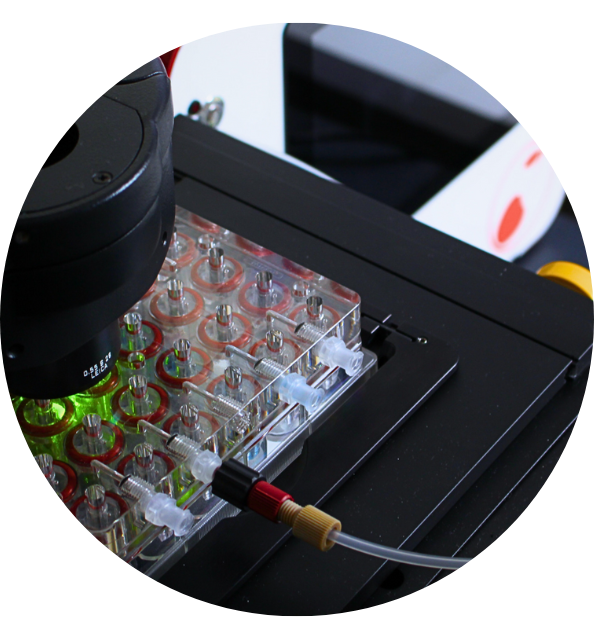
Introduction The researchers created a microfluidic organ-on-chip solution with integrated electrochemical microsensor arrays enabling compartmentalized matrix-based
Introduction Animal models and two-dimensional (2D) cell cultures frequently fail to replicate human drug metabolism and
Introduction Adipose Tissue is very versatile and essential for studies related to Obesity and Diabetes, however,
Introduction The state of the art for tissues-on-chips using skin cells had been lacking for a
Introduction The authors present a simple and modular Microphysiological system without the use of complex components
Introduction Many Organ-on-Chip platforms are still not robust for all cell types, are not reproducible from
Introduction The gut-liver axis is the bidirectional interaction that exists between the gut and its microbiota,
Introduction In vitro generation of perfusable 3D microvessels is an important goal for tissue engineering and
14 rue de la Beaune,
93100 Montreuil (Paris)
France
+33 9 87 04 70 35
contact@cherrybiotech.com
