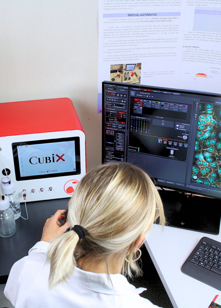
By offering a simple-to-use device, compatible with long-term in-line data recording and
adjustable microenvironment, the CubiX system and the CherryBiotech team provide a highly
valuable tool to design realistic pre-clinical models for therapeutical purposes, in pair with an
advanced platform for research purposes.
We have been enjoying our collaboration and
certainly look forward for more projects and a stronger integration in the Gustave Roussy
comprehensive cancer center.

Institute of Cancerology Gustave Roussy
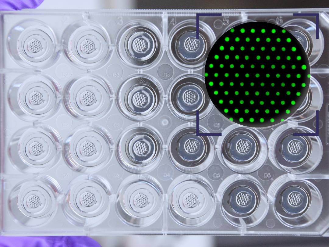
We appreciated having the CubiX in the lab to perform experiments, where we could take advantage of a
6-well flow system. The flow system allows control of the percentages of N2, CO2, and O2 to enrich the
media of the biological samples without needing an incubator. In this system we were able to use disposable transwells, which saved costs for the development of our skin/melanoma -on-chip models, and allowed us to use pre-established protocols, with little adjustments.

The flexibility and potential for customization of the CubiX system is of great benefit for our experiments and ongoing work
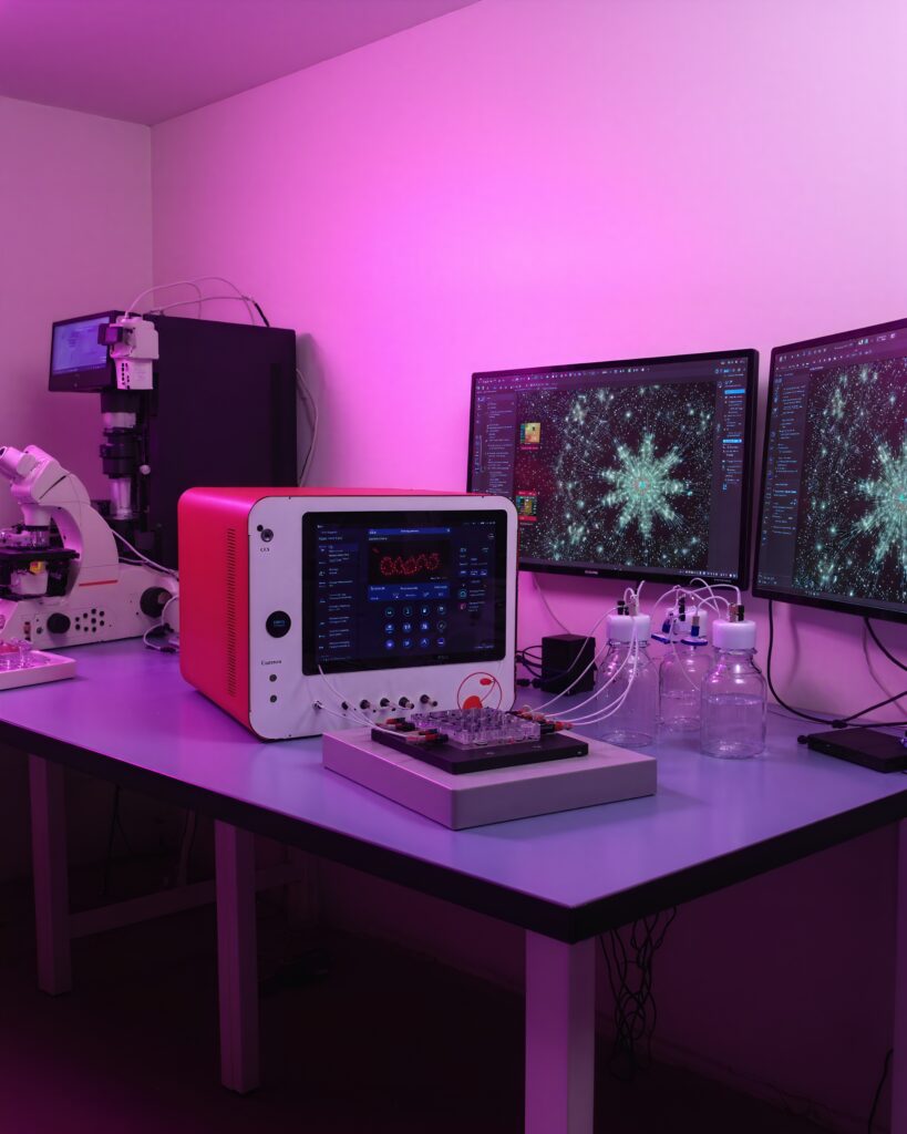
Our experience with the Cubix® prototype provided by Cherry Biotech has been positive. The
support team was exceptionally responsive and helpful, making the process smooth and efficient. While the overall functionality of the bioreactor is impressive, we did encounter some challenges with
troubleshooting minor issues and assembling the tubings. Additionally, the software could benefit from
some UI improvements to enhance usability. Despite these minor points , the advantages of the Cubix®
system are invaluable to our lab in advancing our research on liver biology, and we are eager to continue
our collaboration with Cherry Biotech in the future