Overview
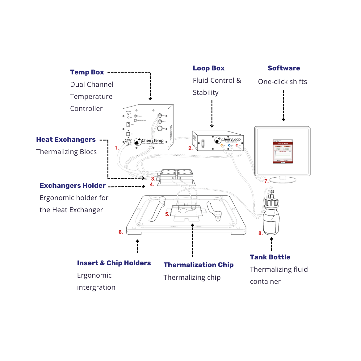
CherryTemp is a temperature controller based on two independent Peltier channels, allowing for very accurate temperature control and ultra-fast shifts from 5°C to 45°C.
A thermalization liquid circulates from a reservoir to the heat exchangers (heating/cooling Peltier elements) where it is thermalized to a target temperature. Once thermalized, this liquid can be quickly injected to a microfluidic device (CherryTemp chip) mounted near the sample. Because of the small thermal mass (the distance between the microfluidic chip and the sample), the temperature transfer is very fast (less than 10 seconds) and of unprecedented accuracy due to a feedback loop control of the room temperature and the heat-sink played by immersion objective lenses.
Specifications
User Interface
Cherry Temp’s user interface offers several advantages, including:
- One-click setting
- Optimized window for easy integration with other software
- Simple navigation
These features make it simple and efficient for users to set up and operate the system, streamlining the experimental process and enhancing productivity.
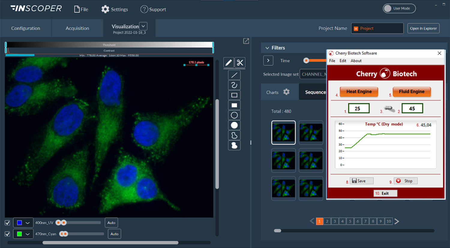
Image: CherryTemp’s interface compatible with INSCOPER software (note: image use authorized by owners)
FAQ
Recent publications feature CherryTemp
Some significative C. elegans articles :
- Dumont et al., 2023, Kinetochore component function in C. elegans oocytes revealed by 4D tracking of holocentric chromosomes, Nature Communications
- Glauser et al., 2023, Multisite regulation integrates multimodal context in sensory circuits to control persistent behavioral states in C. elegans, Nature Communications
- Bowerman et al., 2021, A genetic screen for temperature-sensitive morphogenesis-defective Caenorhabditis elegans mutants, G3
- Seydoux et al., 2019, A gel phase promotes condensation of liquid P granules in Caenorhabditis elegans embryos, Nature Structural & Molecular Biology
- Overholtzer et al., 2019, Entosis Controls a Developmental Cell Clearance in C. elegans, Cell Reports
More C. elegans article published with our CherryTemp system: Read more
Some significative phase separation article:
- Elbaum-Garfinkle et al., 2022, Liquid to solid transition of elastin condensates, PNAS
- Elbaum-Garfinkle et al., 2020, Tunable multiphase dynamics of arginine and lysine liquid condensates, Nature Communications
- Seydoux et al., 2019, A gel phase promotes condensation of liquid P granules in Caenorhabditis elegans embryos, Nature Structural & Molecular Biology
- Kimble et al., 2019, Dynamics of Notch-Dependent Transcriptional Bursting in Its Native Context, Developmental Cell
- Rosen et al., 2018, Nuclear Import Receptor Inhibits Phase Separation of FUS through Binding to Multiple Sites, Cell
More Phase separation articles published with our CherryTemp system: Read more

“The Cherry system is now in full use in my lab and it is working beautifully on its own. The Cherry system is great for stably maintaining temperature on our microscopes”
Dr. Julie Canman
“CherryTemp is a one-of-a kind device for the study of thermosensitive mutants and cytoskeleton dynamics. Its flexibility makes it very easy to adapt to various geometries, from cells to tissue explants or even whole animals !”
Dr. Emmanuel Derivery
“CherryTemp rapidly became one of our favorite piece of equipment in the lab. It’s easy to use and works fast and well. The Cherry Biotech team is always super helpful.”
Dr. Julien Dumont
“The Cherry Temp is perfect for use in imaging core facilities. Its design allows its installation on a wide panel of upright and inverted microscopes and easy training of users thus, promoting development of innovating research project. The customer service is always helpful and responsive.”
Dr. Stéphanie Dutertre
“CherryTemp allows us to perform temperature shift experiments with unprecedented speed and precision with C. elegans embryos. It is very well designed and easy to use. Moreover, we found the the customer service at Cherry Biotech to be very responsive and competent.”
Prof. Pierre Gönczy
“CherryTemp was the perfect solution for us to combine precisely controlled temperature stimuli and live cell imaging in our functional studies on thermosensory neurons in C. elegans adult animals. The system is straightforward to use and we were impressed by the responsiveness of the Cherry Biotech customer service team and by the quality of the support provided.”
Prof. Dominique Glauser


" The Cherry Temp system was essential to acquiring some of our key data on the temperature-dependence of FUS phase separation. The system worked extremely well in these biochemical analyses. It was easy to use and robust, and required only small amounts of material. ”
Prof. Michael Rosen UT SOUTHWESTERN, USA