CHERRY360 – an imaging platform compatible with high-resolution live-cell imaging and yeast synthetic approaches
The Cherry360 program aimed to develop an imaging platform compatible with high-resolution live-cell imaging and yeast synthetic approaches. This projects reflects Cherry Biotech’s story and its deep rootedness in research labs. PI Damien Coudreuse met CEO-founder Jeremy Cramer while the later was developing a cutting-edge microfluidic heater/cooler for microscopy at the Institut Curie, Paris in 2013. Both realized that combining advanced technologies (especially microfluidics) to fundamental biology questions (especially the synthetic biology and the genome duplication and maintenance topics studied by Damien Coudreuse and Jenny Wu) could lead to breaking findings in the field. Supported by the French Brittany Region and ID2Santé, they co-designed the Cherry360 program.
INFO
SKILLS
FIELD OF APPLICATIONS
CELL CULTURE
LONG TERM EXPERIMENTS
FUNDAMENTAL RESEARCH
PUBLICATIONS
A drug-compatible and temperature-controlled microfluidic device for live-cell imaging.
Chen T, Gomez-Escoda B, Munoz-Garcia J, Babic J, Griscom L, Wu PY, Coudreuse D.
LEARN MORE (PDF) – Open Biol. 2016 Aug.


CONSORTIUM
Dr. Jérémy Cramer
CHERRY BIOTECH | FOUNDER & CEO
Julien Babic
PhD student
Dr. Jenny Wu
GDM | GROUP LEADER
Dr. Damien Coudreuse
SYNTHECELL | GROUP LEADER
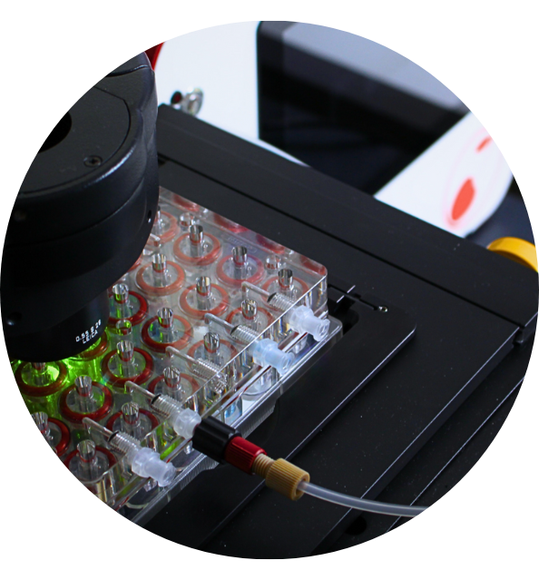
Together with Damien Coudreuse, Cherry co-supervised Julien Babic’s Ph.D. work. In a unique environment offering microfabrication facilities, cutting-edge biological equipment and instrument manufacturing expertise, Julien could develop a microfluidic platform allowing long-time growth and observation of S. pombe cells. Those systems allow both temperature control (including rapid shifts for TS mutant analyses) and medium perfusion (including rapid medium changes) while live-imaging cells with a confocal microscope.
This project has received funding from the French Brittany Region and support from ID2Santé.
