Abstract
We developed a temperature controller that meets cell biology studies requirements. It reaches below ambient temperatures for efficient microtubules depolymerization or endocytosis arrest, it reaches dynamic biology properties by shifting from 5 to 45C in 10 seconds and thus prevents gradient formation inside the sample, it uses electronic control for steady temperature control and microfluidic technology to prevent fluid-flow shear stress on biological samples, it fits on any microscope setting.
Data presented here have been published by Velve-Casquillas et al., 2010 in Lab on a Chip.

Ultra fast temperature shift device for in vitro experiments under microscopy
Introduction
The use of fission yeast temperature-sensitive mutants has contributed to the discovery of new genes involved in yeast biology. They also have been used to gain better understanding in essential genes function. Indeed, essential genes have critical role and mutations in those genes lead to lethal phenotypes, thus preventing further insights on their actions. (Ben Aroya et al., 2010).
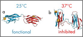
Temperature sensitive mutations are often missense mutations. Mutated proteins retain their functionality at low, permissive, temperature but at higher, restrictive temperature, they become inactive. Fission yeast biology is intrinsically sensitive to temperature. Furthermore, at low temperatures microtubules depolymerize. Here, we present the validation of CherryTemp use for efficient cytoskeleton studies and cell cycle phenotype analysis.
Material and methods
CherryTemp temperature controller device
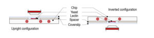
Our PDMS chip is composed of microchannels allowing the circulation of thermalized fluid. The chip is connected to Peltier devices set at two different hot/cold temperatures making possible to quickly change from low to high temperature. Our chip is mounted on a thin glass coverslip,that comes on top of the biological sample, spacers can be used depending on sample thickness. In our system, the fluid is never in contact with the sample thus preventing any shear stress.
Cell imaging and microscopy
Microtubule depolymerization experiments were performed on cdc25-22 expressing GFP-Atb2 tubulin. Mitotic spindle experiments were done using temperature sensitive cut7-24ts expressing GFP-Atb2. Images were acquired using a Yokogawa CSU-10 spinning disc scan head on a Nikon TE2000e inverted microscope. 3D imaging was done using a 100/1.45 NA objective, Metamorph 7.5 software for microscope and Hamamatsu ORCA-AG-CCD camera control.
Results
Reversible ultra-fast cold-induced microtubules depolymerization
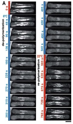
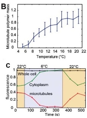
To validate the efficacy of CherryTemp, we used GFP-Atb2 tubulin expressing yeast and follow fluorescence in vivo. At 22C, microtubules exhibit a typical elongated shape (Fig1A). At 16C, we could already quantify a decrease in microtubule polymer mass (Fig1B), and we observed a complete depolymerization when cells were at 6C (Fig1A and 1B) with a loss of microtubule-associated fluorescence signal (Fig1A) and an increase in cytoplasmic-associated fluorescence (Fig1C). This suggests that tubulin dimers were released from the microtubules and move to the pool of free-tubulin dimers. We found that the time to reach full depolymerization was cell-size dependent. This process was reversible, and microtubules repolymerize when we shifted back the temperature from 6C to 22C. Using our temperature controller, we could efficiently monitor cold-induced microtubule depolymerization and re-polymerization while at the microscope stage.
Reversible ultra-fast heat-induced mitosis phenotype
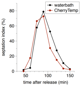
We next wanted to validate the induction of temperature-sensitive mutant phenotype using our device. To this aim, we used Cut7-25ts expressing GFP-Atb2 yeast. Cut7 is a kinesin protein, involved in mitotic spindle formation. At restrictive temperature Cut7-25ts mutant do not form bipolar elongated spindle. At permissive temperature (22C) bipolar spindle is correctly formed and visible with GFP-Atb2 protein (Fig 2A and B). Few minutes after we shifted to 35C, the restrictive temperature, the bipolar spindle retracted and adopted a monopolar shape (Fig2B), this was characterized by a shortening in spindle length (Fig2C). When we shifted the temperature back to permissive level, we could revert the spindle phenotype (Fig2B) and retrieve original spindle length after a few minutes (Fig2C).

References
G. Velve Casquillas, C. Fu, M Le Berre, J. Cramer, S. Meance, A. Plecis, D. Baigl, JJ. Greffet, Y. Chen, M. Piel and PT. Tran Fast microfluidic temperature control for high resolution live cell imaging, Lab on a chip 2010
S. Ben-Aroya, X. Pan, JD. Boeke, and P. Hieter, Making temperature-sensitive mutants, Methods Enzymol. 2010