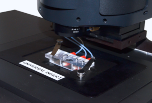
Experimental conditions
Drosophila brains expressing a centriole marker (GFP-PACT) and a microtubule marker (Jupiter-mCherry) were dissected from third instar larvae in PBS and placed in a 10ml drop of Live Cell Imaging Solution (Life Technologies, A14291DJ) on top of the fluidic chamber.
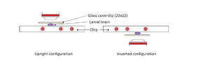
A 22x22mm coverslip was carefully lowered such that the brain became semi-squashed. Filter paper was used to suck excess liquid from the side of the coverslip until a reasonable number of cells had been pushed into a single layer at the edge of the brain.
The sample was initially held at 20°C and then rapidly switched to 5°C.
Microtubule depolymerisation was observed for 370 seconds before the sample was rapidly taken back to 20°C to observe microtubule repolymerisation.
This process was then repeated, with the sample held at 5°C for 260 seconds. All images were processed using Fiji (ImageJ).
Discover the CherryTemp - ultra-fast temperature controler for live imaging in microscopy
Imaging
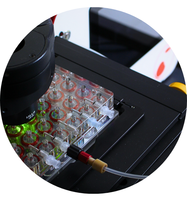
The fluidic slide was then attached to the CherryTemp system and mounted on a Leica DM IL LED inverted microscope controlled by μManager software and coupled to a RetigaR1 monochrome camera (QImaging) and a CoolLED pE-300 Ultra light source using.
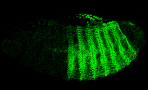
Cells were identified that had centromes arganising robust microtubule asters. Images were taken every 10 seconds using a 63X 1.3NA oil objective (Leica 11506384).
Images were taken of just the Jupiter-mCherry signal due to issues with photobleaching of the GCP-PACT signal.
References
- Courtesy of Paul T. Conduit lab, University of Cambridge, UK.