 Protocol For Drosophila Pupa...
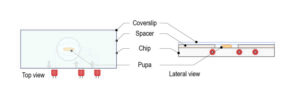
Mounting method for Drosophila pupa With this protocol, spacers of multiple thicknesses can be used to adjust to the diameter of the pupa. S...
Read more Imaging Drosophila Third Instar Larval Brain Cells Expressing Jup...
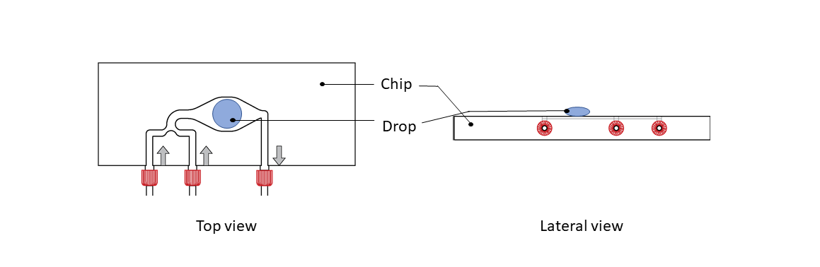
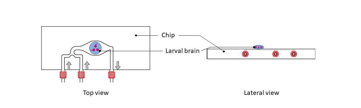
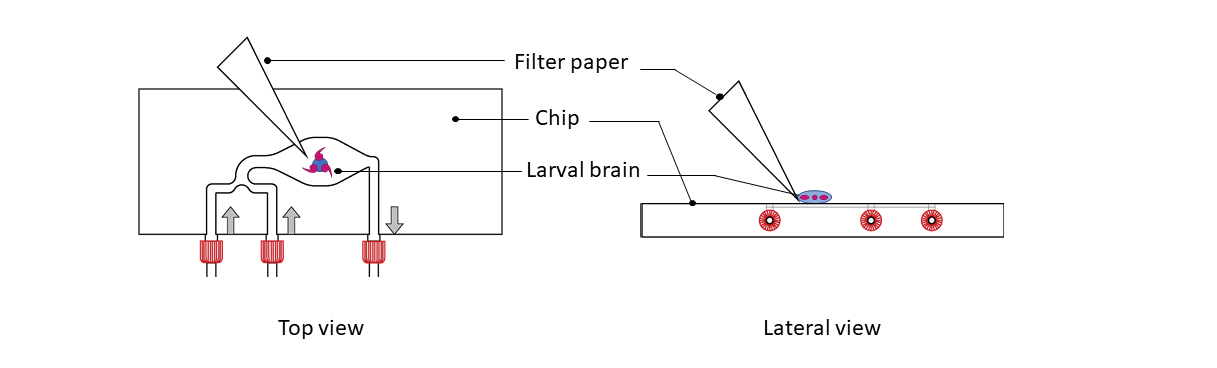
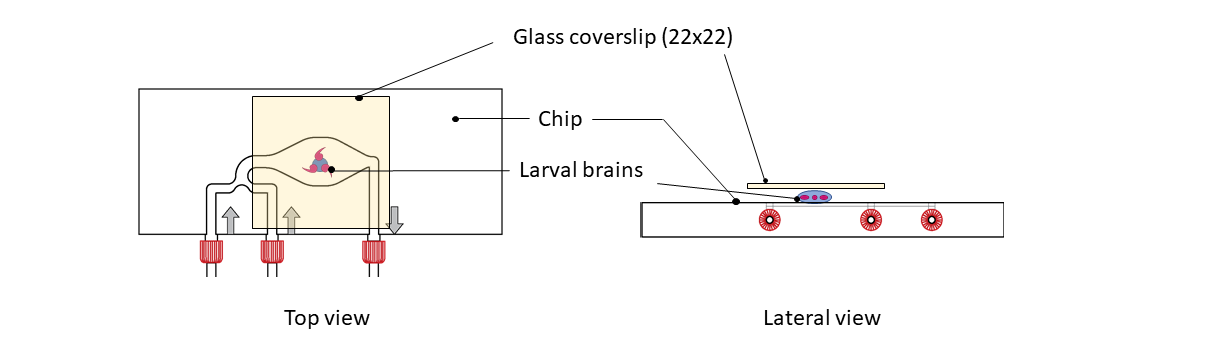
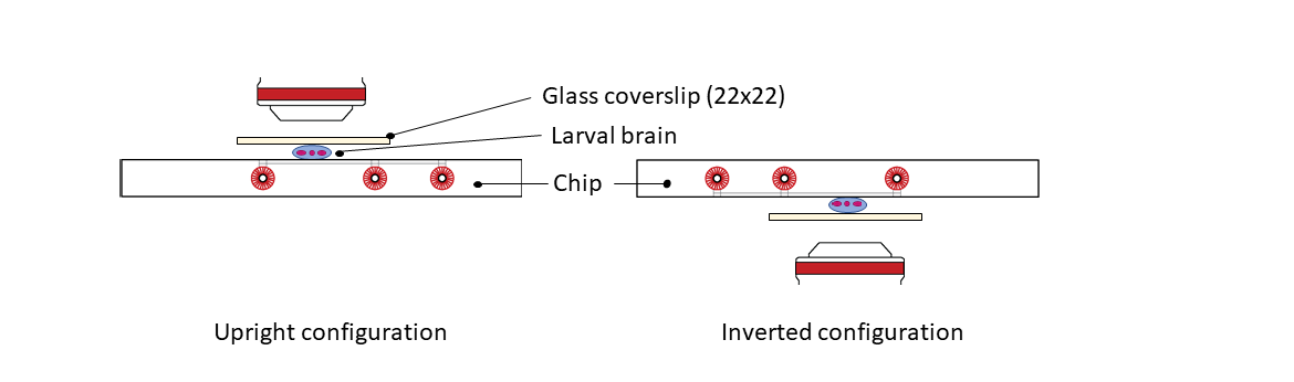
Experimental conditions Drosophila brains expressing a centriole marker (GFP-PACT) and a microtubule marker (Jupiter-mCherry) were dissected...
Read more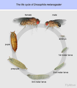 Drosophila Life Cycle And Fly Anatomy...
Introduction The use of D. melanogaster as a model organism in developmental biology has been an important tool to understand fundamental bi...
Read more