Experimental conditions
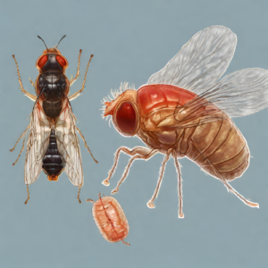
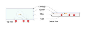
Drosophila third instar larval brains expressing a microtubule marker (Jupiter-mCherry) were mounted on a CherryTemp slide.
The sample was initially held at 20°C and then rapidly switched to 5°C.
Microtubule depolymerisation was observed for 370 seconds before the sample was rapidly taken back to 20°C to observe microtubule repolymerisation. This process was then repeated, with the sample held at 5°C for 260 seconds.
Ultra fast temperature shift device for in vitro experiments under microscopy
Imaging
Cells were imaged on a Leica DM IL LED inverted microscope controlled by μManager software and coupled to a RetigaR1 monochrome camera (QImaging) and a CoolLED pE-300 Ultra light source.
Cells were identified that had centromesorganising robust microtubule asters.
Images were taken every 10 seconds using a 63X 1.3NA oil objective (Leica 11506384).All images were processed using Fiji (ImageJ).
References
Courtesy of Paul T. Conduit lab, University of Cambridge, UK.