 Drosophila: The Fruit Fly Model...
Introduction The fruit fly Drosophila melanogaster (D. melanogaster) has been widely used as a model organism in biological research, partic...
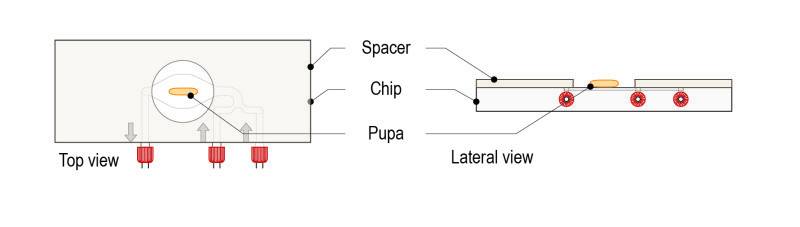
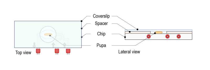
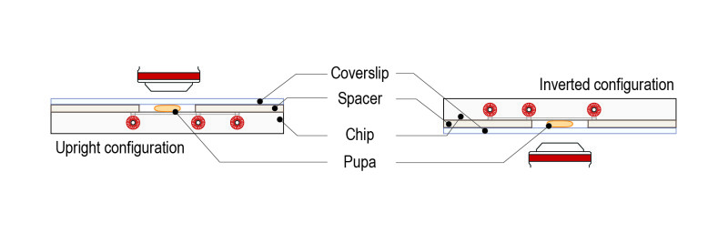
Read more Protocol for Drosophila Larva...
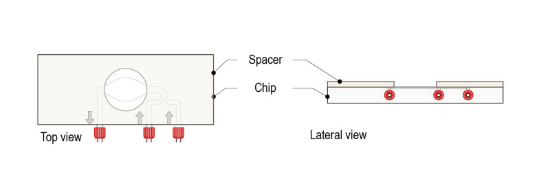
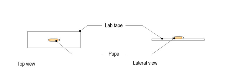
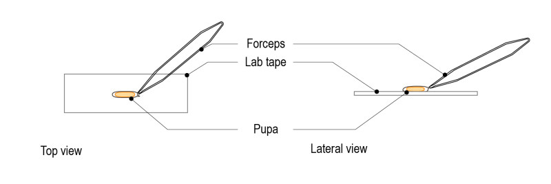
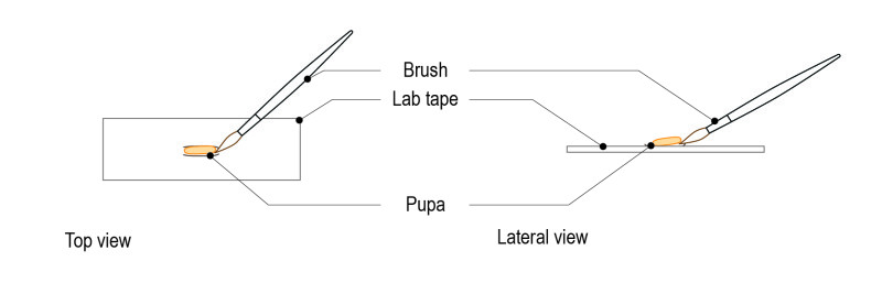
Mounting method for Drosophila larva With this protocol, spacers of multiple thicknesses can be used to adjust to 1st, 2nd and 3d instar lar...
Read more Ion channels experiment...
Temperature control for thermo-sensitive ion channels experiments To sense cold and hot temperatures is vital for all organisms in order to ...
Read more