Embryogenesis in a dish:
Thanks to the last discoveries, a new path has been opened in the human embryo culture and embryonic stem cell culture. Until now, growing in vitro mouse or human embryos were technically limited to cultures that only reached blastocyst stage (5 or 6 days development). The main events in the embryogenesis, especially the ones related to the developmental disorders, occurs just right after those days (1), highlighting the need of having access to older stage embryos.
Since 2016, it is possible to culture human embryos up to 13 days because of technical improvements. This is allowing us to observe many of the key structures and events that will later support the growth of the embryo. Indeed, after 14 cell cycles, the embryo is preparing itself to undergo the gastrulation, which is the main organizational event that generates the basic body plan and provides a base to build all the tissues (2-3).
 Figure 1: A scheme for the human embryogenesis process from the first division until the birth.

Discover Cubix
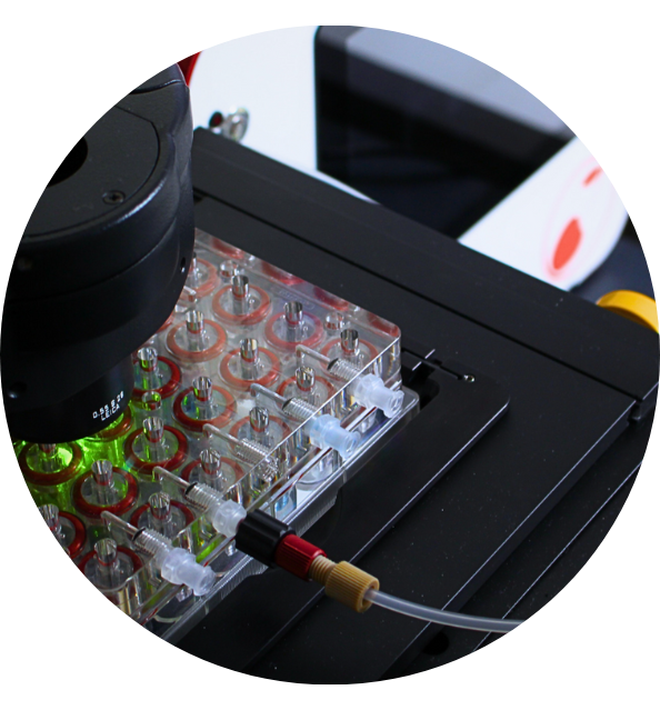
The sum of the next-generation 3D cells culture technologies.
The ability of embryonic stem cells to divided and be renewed make them a good and easily handling experimental model. Furthermore, and by definition, they can become any types of cells after going through many key stages of development. While the in vivo embryogenesis is well spatially organized, in vitro differentiation of embryonic stem cells is not. The possibility for researchers to engineer the microenvironment to make it suitable for the three-dimensional tissue differentiation is a key advance. It is now possible to provide devices and structures that closely mimic the shape of a differentiating embryo (4-5). Going in that way, there are some recommendations from Harrison and colleagues (6) that could led us to the final goal of mimicking human embryogenesis in a dish. One of the main drawback is that the supply of human embryos is limited and very few of these embryos reach advanced developmental stages. Because extra-embryonic tissues (placenta or yolk sac) provide, in addition to nutrients, signals that drive the differentiation of pluripotent cells in a regulated spatial and temporal domain, combining embryonic stem cells with trophoblast stem cells might be a solution. Indeed, the combination of these two types of cells in a gel containing extracellular matrix molecules produces promising results in mouse.
Even if these replicas of mouse embryos were not perfect they represent a first step on the race to make embryogenesis on a dish. The key now is to understand how refining the culture environment (spatial, chemical and physical) for the embryos to develop. To do so, a multidisciplinary research will be required: e.g biologist will need help from biochemist and engineer to develop a scaffold for the embryos to growth.
Bibliography/Sources
- Pera M. Embryogenesis in a dish. Science
- Deglincerti et al., Nature 533, 251 (2016).
- N. Shahbazi et al., Nat. Cell Biol. 18, 700 (2016).
- Etoc et al., Dev. Cell 39, 302 (2016).
- Shao et al., Nat. Mater. 16, 419 (2016).
- E.Harrison, B. Sozen, N. Christodoulou, C. Kyprianou, M. Zernicka-Goetz,Science 356, eaal1810 (2017)
Pablo is part of the H2020-MSCA-ITN-ETN-DivIDe European network. LEARN MORE.

Discover the CubiX
Emulating human cell & tissue physiology


