 Automated Phenotyping Of Caenorhabditis Elegans Embryos...
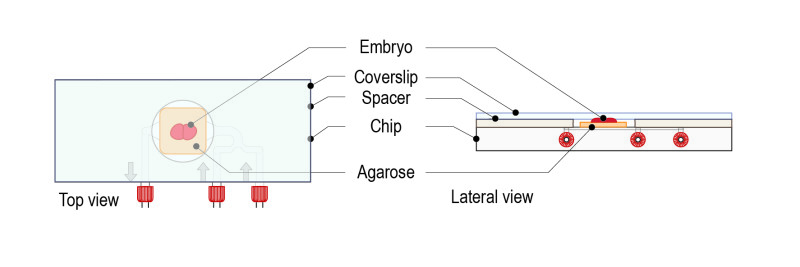
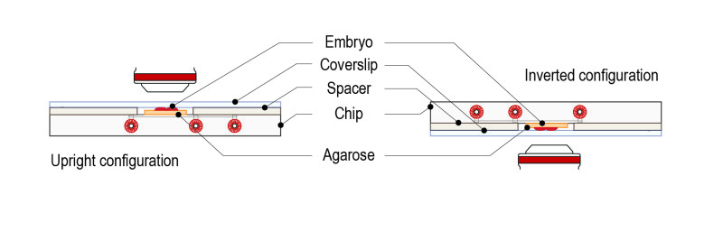
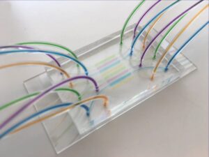
A new microfluidic system allows the high throughput phenotyping of C. elegans embryos! Martin A. M. Gijs et al. produced a high-throughput ...
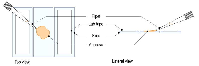
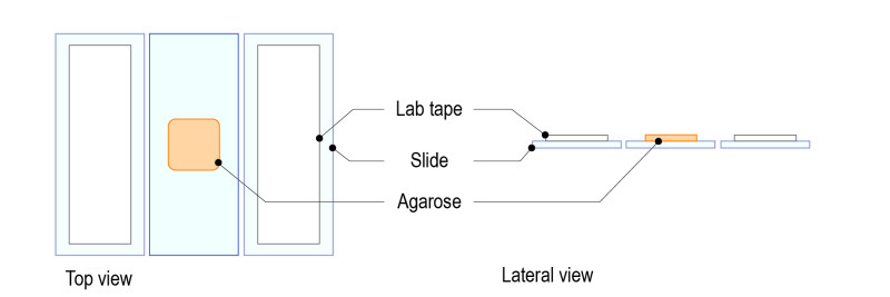
Read more Agar pad for live cell imaging...
Introduction Non-adherent cells are difficult to image because of movement. This is the case of living cells which are motile, but also of f...
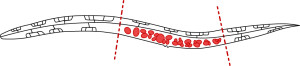
Read more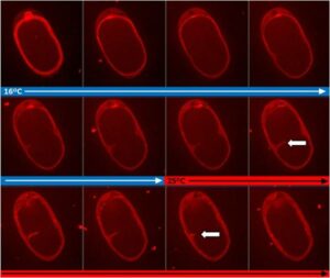 Cytokinesis Arrest in C. elegans Temperature Sensitive Mutants...
This application note shows how cytokinesis can be perturbed using CherryTemp, by a rapid temperature shift of a C. elegans CYK-4 thermose...
Read more