Temperature control of mammalian cell metabolism
In Human cells, regulation of body temperature and adaptation to temperature change is a major physiological function. At the cellular level, cells respond to a temperature decrease by decreasing metabolism, membrane fluidity, transcriptional activity, protein synthesis and vesicular transport rates. Mitochondria are the center of cell metabolism and play crucial role in regulating and maintaining cells temperature.
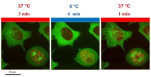
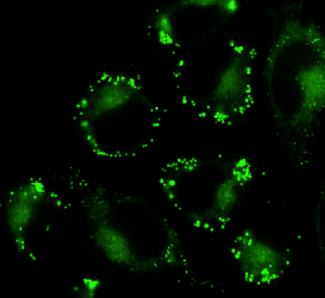
With CherryTemp, you can tightly control and ultra-quickly shift temperature, from 5 to 45°C in less than 10 seconds, while imaging mitochondrial protein immediate response to intracellular temperature change, take a tour!
Mitochondria metabolism
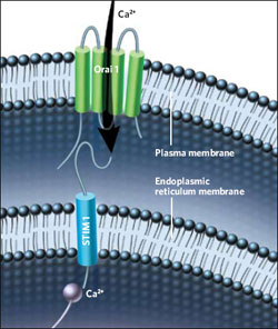
Mitochondria are specialized organelles and are the metabolic center of the cell. They play a role in crucial biochemical pathways generating chemical energy for the cells. They are involved in cell death pathways, phospholipid metabolism, calcium sensing. In pancreatic beta-cells, mitochondria couple glucose metabolism to insulin release. Upon glucose metabolism, mitochondria produce ATP which binds and close K+ channel, leading to Ca2+ ion channel opening, Ca2+ entry into the cell leads to insulin vesicles release. Mitochondria are very sensitive to temperature, and in particular they participate in cellular thermogenesis.
Ultra fast temperature shift device for in vitro experiments under microscopy
Cold induced thermogenesis
In mammals, brown adipose tissue is involved in the maintenance of body temperature. Brown adipocytes have a common embryonic origin with skeletal muscle and while brown fat tissue is abundant at birth and decreases with age. When temperature is low, sympathetic nerves stimulate brown adipocytes by realeasing norepinethrin, which in turn binds on beta-adrenergic receptors abundantly expressed at the brown adipocytes cell surface. Stimulation by norepinephrin triggers a signaling cascade leading to the rapid activation of UCP-1, an uncoupling protein playing an important role in the mitochondrial adaptative response to cold. Ucp-1 is localized at the mitochondrial inner membrane and it acts by uncoupling proton gradient from ATP production at the benefit of heat production. UCP1 is Paramount for cold-induced thermogenesis in brown adipocytes. UCP-1 is specific of Brown adipocyte. UCP2 is ubiquitously expressed, UCP3 is highly expressed in skeletal muscle cells mitochondria.

Cellular metabolism imaging
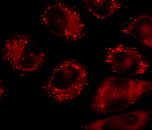
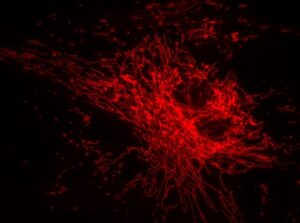
Advances in microscopy techniques and the availability of specific mitochondrial fluorescent markers allowed scientists to improve their knowledge in mitochondrial biology. Mitra and Lippincott-Schwartz have reviewed the different imaging technique to track mitochondria. Choosing the adequate fluorescent markers is of importance and depends on the type of experiments. Since mitochondria are very sensitive to cell stress, it is best to avoid transient transfection of fluorescent proteins, a source of stress for the cells, and rather establish stable cell lines with a fluorescent version of the protein of interest. If the experiments require long-term imaging, it is important to choose a fluorescent marker whose wavelength is not strongly energetic in order to prevent mitochondrial damage. Very good mitochondrial markers are mitoRFP, YFP or GFP (Mitra and Lippincott-Schwartz, 2010). For short-term experiment (less than 45min) on live cell, the use of fluorescent dyes which are taken up by mitochondria may prove useful. Mitotracker green is one such fluorescent dyes, it is absorbed by mitochondria regardless of its membrane potential.
Combine live-cell imaging and temperature shift experiments
Mitochondria are dynamic organelles, they can change their shape from filamentous to granular structures, they move inside the cells and are transported along microtubules. Mitochondria are the cell metabolic center, and it is clear that metabolic pathways are highly sensitive to temperature. Temperature changes affect biosynthesis and degradation, and the rate of enzymatic reaction. Notably cancer cells, which keep on dividing, have higher internal temperature, likely due to increased metabolic rate. It is therefore very important to understand how temperature regulate cellular metabolism. Imaging techniques have greatly developed to tackle mitochondria biology with live-cell imaging.
Now you can benefit from CherryTemp to study the dynamic relationship between temperature and cell metabolism. CherryTemp allows you to change temperature from 5-45°C in less than 10 seconds, the system is easy to use and unlike other system, it does not require the use of an additionnal objective heater collar.
References
Zohar O, Ikeda M,Shinagawa H,Inoue H,Nakamura H,Elbaum D, Alkon DL, and Yoshioka T,Thermal Imaging of Receptor-Activated Heat Production in Single Cells, Biophysical journal, 1998
Kong X, Banks A, Liu T, Kazak L , Rao RR, Cohen P, Wang X , Yu S , Lo JC, Tseng YH, Cypess AM, Xue R , Kleiner S, Kang S, Spiegelman BM. , RosenED. IRF4 Is a Key Thermogenic Transcriptional Partner of PGC-1α, Cell, 2014
Rousset S , Alves-Guerra MC , Mozo J , Miroux B , Cassard-Doulcier AM , Bouillaud F , and Ricquier D, The Biology of Mitochondrial Uncoupling Proteins, Diabetes, 2004
K Mitra and Lippincott-Schwartz J, Analysis of mitochondrial dynamics and functions using imaging approaches Curr Protoc Cell Biol. 2010
See also :
Cellular Metabolism Resources from Cell Signaling
What Is Cellular Metabolism? fromGlobal Healing Center
FAQ
The regulation of body temperature is a major physiological function in humans. At the cellular level, cells react to a temperature decrease with several changes. A general decrease in metabolism is observed. The fluidity of the cell membrane is also lessened. Other cellular processes are slowed. These include transcriptional activity and the rate of protein synthesis. The rate of vesicular transport is also decreased in response to colder conditions. Mitochondria, which are the centre of cell metabolism, are involved in regulating and maintaining the temperature of cells. These organelles are very sensitive to temperature.
Mitochondria are specialised organelles and are known as the metabolic centre of the cell. They are involved in important biochemical pathways that generate chemical energy for the cell. Mitochondria also participate in other processes, such as cell death pathways, phospholipid metabolism, and calcium sensing. Their function is very sensitive to temperature, and they participate in cellular thermogenesis. In pancreatic beta-cells, for instance, mitochondria link glucose metabolism to the release of insulin. The ATP produced by mitochondria binds to and closes K+ channels. This action leads to the opening of Ca2+ ion channels, and the resulting entry of Ca2+ into the cell leads to the release of insulin vesicles.
In mammals, brown adipose tissue is involved in maintaining body temperature. When the temperature is low, sympathetic nerves stimulate brown adipocytes. This stimulation occurs through the release of norepinephrine, which binds to beta-adrenergic receptors on the cell surface. This action starts a signalling cascade. The cascade leads to the rapid activation of UCP-1, an uncoupling protein. UCP-1 is located at the mitochondrial inner membrane. It functions by uncoupling the proton gradient from the production of ATP. This uncoupling results in heat production instead of energy storage. This protein is required for cold-induced thermogenesis in brown adipocytes and is specific to this cell type.
Choosing the correct fluorescent marker is an important consideration. This choice depends on the type of experiment being performed. Mitochondria are known to be very sensitive to cell stress. For this reason, it is best to avoid the transient transfection of fluorescent proteins, as this is a source of stress for the cells. A better method is to establish stable cell lines that express a fluorescent version of the protein of interest. If the experiments require long-term imaging, a fluorescent marker with a wavelength that is not strongly energetic should be chosen. This precaution helps to prevent mito chondrial damage. For short-term experiments on live cells (less than 45 minutes), fluorescent dyes that are taken up by mitochondria may be useful, such as Mitotracker green.