Application notes

Scientific Poster – Skin sensitization and safety assessment based on an in vitro 3D vascularized fully differentiated immunocompetent skin model

SCIENTIFIC POSTER DOWLOAD —
Skin sensitization and safety assessment based on an in vitro 3D vascularized fully differentiated immunocompetent skin model

Fassini D, Mejri H, Fernandes P, Simon M, Homs Corbera A, Poinsot C, Gaudriault P, Filaire E

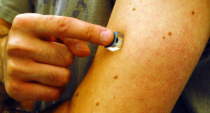
With the rising prevalence of allergic contact dermatitis, accurately assessing the skin sensitization potential of chemical products is increasingly critical. Current methods, guided by three OECD guidelines and a set of in vitro tests, have advanced with skin model equivalents (SMEs). However, these models lack the crucial interaction between epidermis/dermis cells and resident immune cells, a key factor in skin sensitization.

Our study aimed to develop an SME that encompasses an endothelial barrier and relevant resident immune cells, enhancing the understanding of skin sensitization processes. We integrated adult Normal Human Epidermal Keratinocytes, Normal Human Dermal Fibroblasts, Human Umbilical Vein Endothelial Cells, THP-1 cells, and THP-1 derived macrophages into our model.

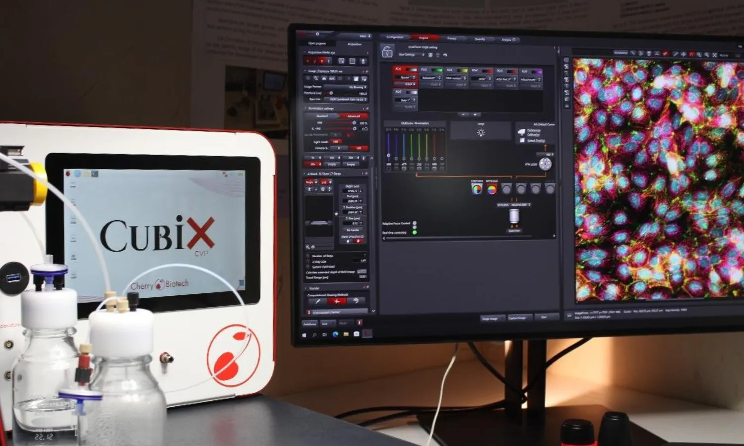
This innovative approach led to a multi-layered, differentiated epidermis that proliferates atop the dermis, as confirmed by immunolabelling analyses. Our model also successfully verified the presence and activation of specific human epidermis/dermis markers and resident immune cell phenotypes, offering a more comprehensive method for skin sensitization and safety assessment.

The poster, title: A New alternative method for skin sensitization and safety assessment based on an in vitro 3D fully differentiated skin model – is free to download here. Don’t miss out!

Cherry Biotech-Poster-Skin-sensitization-in-vitro-3D-differentiated-skin-model

How to culture vascularized & immunocompetent 3D models in a standard Multiwell

Related Posts

Schematic representation of an adipose-tissue-on-chip module illustrating the principal engineering elements that enhance physiological relevance: (i) perfusable lumen with circulating immune cells permitting real-time trafficking; (ii) endothelial monolayer aligned by shear stress to maintain a selective barrier; (iii) continuous perfusion for nutrient delivery and waste removal; (iv) controlled gas exchange enabling oxygen tuning and hypoxia modelling; (v) intrinsic, perfused microvasculature embedded within the stromal-vascular fraction; and (vi) fluidic coupling to a downstream liver compartment to study multi-organ crosstalk. A legend identifies the main cell types and extracellular matrix components.
Adipose Tissue-on-Chip: Perfused Platforms Driving Next-Gen Metab...
Obesity, type 2 diabetes, and non-alcoholic fatty liver disease (NAFLD) remain major global health concerns, yet preclinical models continue...
Read more
Cultivating gut microbiota on a gut-on-a-chip...
A complex human gut microbiome cultured in anaerobic intestine-on-a-chip A recently published paper on Nature biomedical engineering highl...
Read more
3d-cell-culture-Microfluidic-3D-Endothelium-on-a-Chip
A Microfluidic 3D Endothelium-on-a-Chip Model to Study Transendot...
Introduction Microfluidic 3D Endothelium-on-a-Chip – The endothelium is a thin membrane that lines the heart and blood vessels. Endothelia...
Read more

get in touch

Get the best insights about Cherry Biotech by Email Let’s stay in touch!
As part of our commercial prospecting, we may need to process your personal data. For more information, please consult our Privacy Policy