Application notes

Scientific Poster – Development of a 3D immunocompetent breast cancer model to accelerate drug testing and personalised medicine

Authors: Priyanka Fernandes, Harry Dawson, Ethan N.P Vong, Pierre Savagner, Dario Fassini


Breast cancer remains one of the leading causes of cancer-related mortality in women. To address patient heterogeneity, overcome drug resistance, prevent metastasis, and improve treatment efficacy, personalized therapeutic approaches are gaining increasing importance. The tumour microenvironment (TME) plays a critical role in driving cancer progression and shaping therapeutic outcomes. However, most existing 3D in vitro models fail to adequately mimic tumour–stromal complexity.

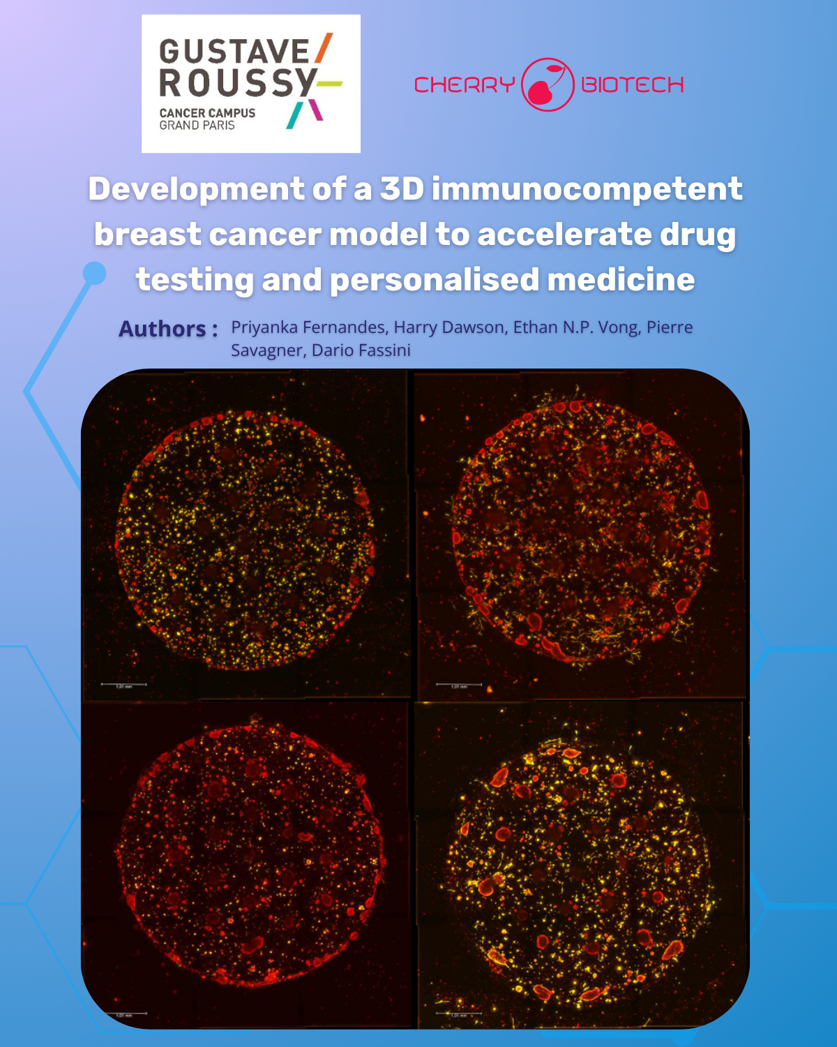
We present our 3D immunocompetent breast cancer model, designed to support personalized medicine and drug screening with a human, physiologically-relevant platform capturing the tumor microenvironment interactions.

  • Luminal B breast cancer model from patient-derived xenograft cells (PDX) from individual resistant to first-line treatment, showcasing the potential for personalized therapy 
  • Fibroblasts and macrophages co-culture in ECM demonstrating the influence of the microenvironment on therapy resistance
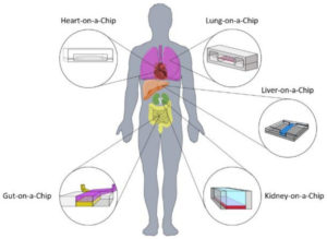
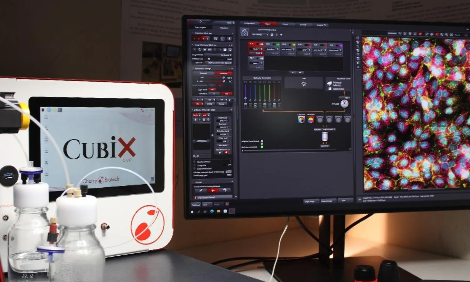
  • Integration of the model into our CubiX microphysiological system enabling real-time monitoring of oxygen, glucose, lactate and temperature
  • Evaluation of of chemotherapies using live imaging, biochemical assays, immunofluorescence and histology

Download our poster to see how our breast cancer model powered by our technology is supporting precision medicine and oncology research. 

The poster, titled: Development of a 3D immunocompetent breast cancer model to accelerate drug testing and personalised medicine – is free to download here. Don’t miss out!

breast-cancer-poster-mps-brussels-webpage

How to culture vascularized & immunocompetent 3D models in a standard Multiwell

Related Posts

Cartoon-style illustration of scientific papers about adipose tissue and metabolism, surrounded by icons of fat cells, molecules, and research graphs.
10 Groundbreaking Studies Using 3D Adipose Tissue Models (2020-20...
Obesity and its related metabolic diseases, such as type 2 diabetes, represent a growing global health crisis, yet progress in treatment has...
Read more
embryogenesis-cult_cell_souches5-smart-servier
How Far Are We From Embryogenesis in a Dish?...
Embryogenesis in a dish Thanks to the last discoveries, a new path has been opened in the human embryo culture and embryonic stem cell cultu...
Read more
Flowchart showing the steps used to create patient-derived tumor organoids (PDTOs): starting at left with a human silhouette and icons for “biological fluids” and “tumor”, followed by enzymatic/mechanical dissociation equipment; a tube containing isolated cells; embedding of cells in an extracellular-matrix dome inside a culture well; addition of enriched medium; formation of PDTOs; parallel branches for characterization (histology slide, sequencing machine), research assays (multi-well plate, radiation symbol, pill), and cryopreservation (vial rack); arrows trace feedback from PDTO characterization back to the original tumor to correlate findings.
Breast cancer organoid: A short history of innovation in breast c...
Breast cancer has long been one of the most prevalent and challenging cancers worldwide, demanding constant advances in treatment and resear...
Read more

get in touch

Get the best insights about Cherry Biotech by Email Let’s stay in touch!
As part of our commercial prospecting, we may need to process your personal data. For more information, please consult our Privacy Policy