Application notes

SCIENTIFIC POSTER — Charting new paths in adipose tissue organoid development via stromal fraction

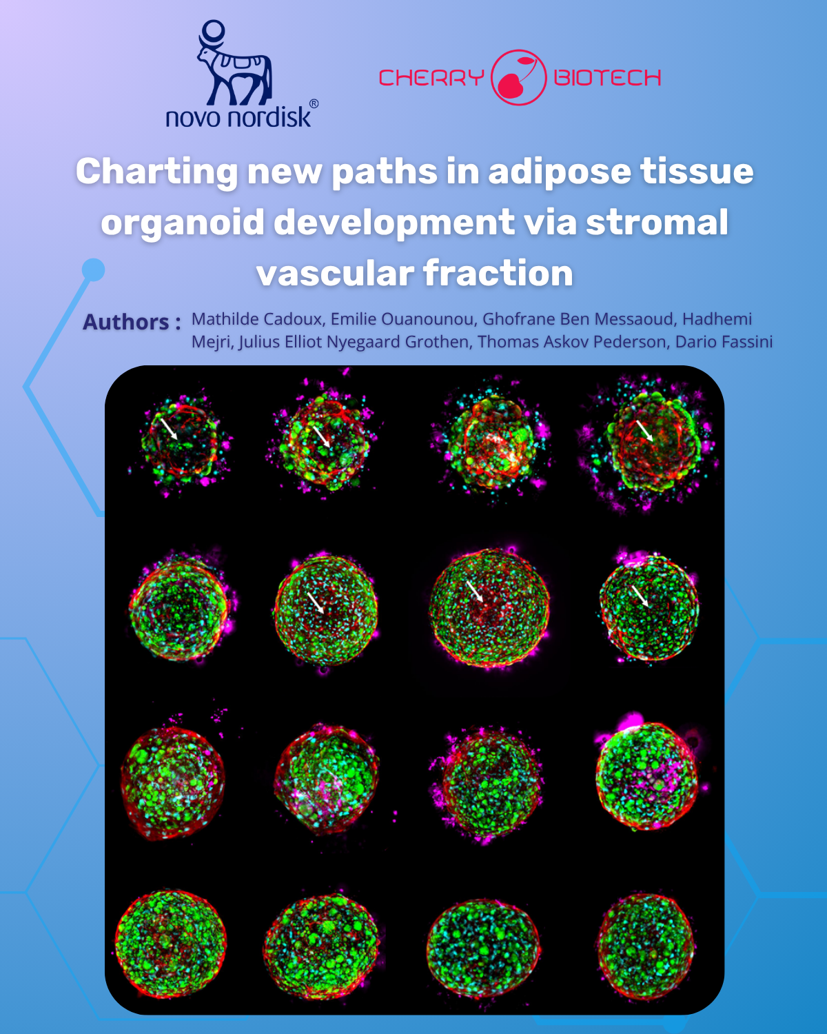
Authors: Mathilde Cadoux, PhD; Emilie Ouanounou; Ghofrane Ben Messaoud; Hadhemi Mejri; Julius Elliot Nyegaard Grothen, PhD; Thomas Askov Pedersen, PhD; Dario Fassini, PhD

Metabolic diseases are strongly linked to adipose tissue dysfunction, yet current in vitro models poorly capture the biology of mature white adipose tissue.

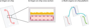
In collaboration with Novo Nordisk, we optimized protocols to generate SVF-derived adipose organoids using Cherry Biotech’s CubiX microphysiological system. Our work focused on:

  • Developing SVF-derived
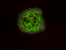
    organoids reproducing key features of white adipose tissue, including
    unilocular adipocytes and endothelial cells

  • Exploring perfusion and
    hydrogel effects on adipose organoid morphology, viability, and
    differentiation

  • Validating cryopreserved SVF as
    a robust source, enabling reproducible and physiologically relevant white adipose tissue models

Download our poster to discover how this model supports metabolic disease research and drug discovery.

In this study, CubiX has enabled precise control of oxygen, nutrients, and flow, supporting the advancement of more refined and reliable in vitro organoid models.

The poster, titled: Charting new paths in adipose tissue organoid development via stromal vascular fraction – is free to download here. Don’t miss out!

Poster featuring fluorescent microscopy images of human adipose tissue organoids derived from stromal vascular fraction (SVF). The images display spherical 3D organoids with green, red, and magenta fluorescence highlighting different cell populations and extracellular structures. Logos of Novo Nordisk and Cherry Biotech appear at the top, with the title “Charting new paths in adipose tissue organoid development via stromal vascular fraction” and a list of contributing authors.

SCIENTIFIC POSTER — Charting new paths in adipose tissue organoid development via stromal fraction

How to culture vascularized & immunocompetent 3D models in a standard Multiwell

Related Posts

Poster cover with Gustave Roussy and Cherry Biotech logos, title ‘Development of a 3D immunocompetent breast cancer model to accelerate drug testing and personalised medicine,’ authors listed, and four fluorescent microscopy images of circular tumor spheroids with red/yellow immune-cell staining on a blue gradient background.
Scientific Poster – Development of a 3D immunocompetent breast ...
Authors: Priyanka Fernandes, Harry Dawson, Ethan N.P Vong, Pierre Savagner, Dario Fassini   Breast cancer remains one of the leading causes...
Read more
Skeletal Muscle Biopsy Beyond Histological Diagnosis: Ex-vivo Cul...
Skeletal Muscle: Overview, Histology, and Pathology Muscle tissue represents one of the four tissue in the human body with the epithelial ti...
Read more
Illustration of human liver tissue showing classical lobule structure with central vein and portal triads, stained with hematoxylin and eosin
In vitro liver models in drug discovery: Main models, advantages ...
In vitro liver models are central to drug discovery and toxicology, enabling controlled evaluation of metabolism, disposition, and safety be...
Read more

get in touch

Get the best insights about Cherry Biotech by Email Let’s stay in touch!
As part of our commercial prospecting, we may need to process your personal data. For more information, please consult our Privacy Policy