Overcoming the limit of light diffraction in microscopy
Light diffraction is a physical phenomenon that define the resolution limits in both optical and electron microscopy. This phenomenon has been discovered and characterized by Abbe in 1873; he found that the capability to resolve two points at a certain distance is related to the wavelength used to illuminate the object. In the last decade new light microscopy techniques based on signal processing and/or light beam manipulation have made possible to go beyond the limits of diffraction.
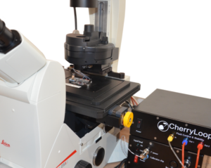
Ultra fast temperature shift device for in vitro experiments under microscopy
Diffraction in optical microscopy (super resolution microscopy)
Optical microscopy, also referred as light microscopy, is the oldest technique of microscopy. It is based on a light source of visible light to illuminate a sample, light passing trough the sample is then collected and magnified by a group of lenses (objective and ocular). The same principle is still used nowadays and is called bright field microscopy. It is still a useful technique for routine analysis and fast screenings in life and material science.
The first basic optical/light microscopes were developed during the 17th century. Nowadays, several type of microscopy techniques have been developed in order to allows scientist to obtain different information of their specimens.
There are many factors that can affect the final resolution of an optical imaging system like a microscope but the most relevant and difficult to overcome is the diffraction limit of light given the fact that it is a physical law and does not depends on the quality of the lenses and the way the optics are arranged.
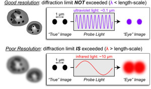
Back in 1873, the German physicist Ernst Abbe discovered how the resolving power of microscopes is limited by the diffraction of light. His research revealed that the resolution of a microscope is not related with the quality of the microscopy components themselves but due to the wavelength of the light used and the aperture of the optics setup.
Due to this phenomenon, a microscope is not able to resolve two objects if they are located to a distance that is shorter that λ/2NA, with λ being the wavelength of light used to illuminate the sample and NA the numerical aperture of the imaging lens. This implies that the diffraction happening at the sample level does not allow to differentiate between two objects divided by a lateral distance which is less than half of the wavelength of the light that has been used to image the sample [2-3].
In other words the resolution of a microscope is inversely proportional to the wavelength of the light that is observed and directly proportional to the size of the objective itself.
Abbe discovered that images are made of an array of diffraction-limited spots with different intensities which overlap one to each other to produce the final image. While it is possible to increase the resolution by reducing the wavelengths (i.e. using UV light) or by increasing the numerical aperture it is not possible to go beyond a resolution of 200nm [4].
Nevertheless in the last decades advances in fluorescent microscopy, light source manipulation and image signal processing had led to the development of several microscopy techniques able to overcome the physical constrain of the Abbe law. Those new type of microscopes are known as Super Resolution microscopy [5].
References
- Beyond the diffraction limit. Nat Photonics [Internet]. 2009 Jul 1 [cited 2017 Jun 15];3(7):361–361. Available from: http://www.nature.com/doifinder/10.1038/nphoton.2009.100
- The Diffraction Limits in Optical Microscopy [Internet]. [cited 2017 Jun 15]. Available from: http://www.azooptics.com/Article.aspx?ArticleID=659
- The Diffraction Barrier in Optical Microscopy | MicroscopyU [Internet]. [cited 2017 Jun 15]. Available from: https://www.microscopyu.com/techniques/super-resolution/the-diffraction-barrier-in-optical-microscopy
- Riedl MJ, J. M. Optical design fundamentals for infrared systems [Internet]. Optical design fundamentals for infrared systems, 2nd ed. Bellingham, WA: SPIE, The International Society for Optical Engineering, 2001, xvii, 182 p. SPIE tutorial texts TT 48, ISBN 0819440515. SPIE Press; 2001 [cited 2017 Jun 15]. 182 p. Available from: http://adsabs.harvard.edu/abs/2001odfi.book…..R
- Super-resolution microscopy contents from Nature : https://www.nature.com/collections/vvfdnjgfrj
FAQ
Light diffraction is a physical phenomenon that defines the resolution limits for optical microscopy. It was characterized by Abbe in 1873\. He found that the ability to resolve two points is related to the wavelength used for illumination. Due to this phenomenon, a microscope cannot resolve two objects if they are at a distance shorter than λ/2NA. The variable λ is the wavelength of light, and NA is the numerical aperture of the lens. This means objects with a lateral distance less than half the light’s wavelength cannot be differentiated. This is a physical law, not a limit based on the quality of the optics. For optical microscopes, it is not possible to go beyond a resolution of 200nm using traditional methods.
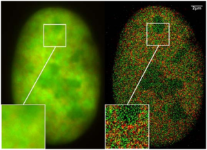
In the last decades, new light microscopy techniques have made it possible to go beyond the limits of diffraction. These developments are based on advances in fluorescent microscopy. They are also based on light source manipulation and image signal processing. These new methods are able to overcome the physical constraint of the Abbe law, which was first described in 1873\. Microscopes that use these new techniques are known as Super-Resolution microscopy. A comparison shows that what appears as a homogeneous and blurry signal in standard fluorescence microscopy can be turned into a detailed map of the signal when these newer methods are used.
In 1873, the German physicist Ernst Abbe discovered how the resolving power of microscopes is limited by the diffraction of light. His research showed that the resolution of a microscope is not related to the quality of the microscopy components themselves. Instead, it is due to the wavelength of the light being used and the aperture of the optics setup. He found that the capability to resolve two points at a certain distance is related to the wavelength used to illuminate the object. Abbe also discovered that images are made of an array of diffraction-limited spots. These spots have different intensities and overlap one to another to produce the final image.
Optical microscopy, which is also referred to as light microscopy, is the oldest microscopy technique. The first basic light microscopes were developed during the 17th century. The principle is based on a light source of visible light, which is used to illuminate a sample. The light that passes through the sample is then collected. After collection, it is magnified by a group of lenses, specifically the objective and the ocular. This same principle is still used in the present day. This method is called bright field microscopy. It remains a useful technique for routine analysis and for fast screenings in both life and material science.