Introduction
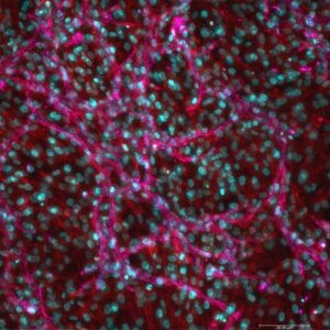
In postmenopausal women, fat mass and adipose-tissue mass (see Human Breast Adipocytes) are closely linked to obesity and breast cancer. Adipocytes have the ability to modulate the tumor microenvironment by secreting energy nutrients, which increases the risk of breast cancer incidence, proliferation, and metastasis.
Physical and emotional repercussions are significantly linked to cancer incidence and growth, in addition to other obesity-related issues. As a result, it’s critical to comprehend how adipose tissues and adipocytes can influence breast cancer risk and mortality.
How to culture vascularized & immunocompetent 3D models in a standard Multiwell
Abstract
Background/aim: Recent research highlights the role of cancer-associated adipocytes (CAA) in promoting breast cancer cell migration, invasion and resistance to therapy. This study aimed at identifying cellular proteins differentially regulated in breast cancer cells co-cultured with CAA.
Materials and methods: Adipocytes isolated from human breast adipose tissue were co-cultured with hormone receptor-positive (MCF-7) or -negative (MDA-MB-231) breast cancer cells using a transwell co-culture system. Proteomes of co-cultured and control breast cancer cells were compared quantitatively using iTRAQ labelling and tandem mass spectrometry, and the results were validated by western blotting.
Results: A total of 1,126 and 1,218 proteins were identified in MCF-7 and MDA-MB-231 cells, respectively. Among these, 85 (MCF-7) and 63 (MDA-MB-231) had an average fold change >1.5 following co-culture. Pathway analysis revealed that CAA-induced enrichment of proteins involved in metabolism, the ubiquitin proteasome, and purine synthesis.
Conclusion: This study provides a proteomic platform for investigating the paracrine role of CAA in promoting breast cancer cell metastasis and resistance to therapy.
References
Lee Isla Crake R, Phillips E, Kleffmann T, Currie MJ. Co-culture With Human Breast Adipocytes Differentially Regulates Protein Abundance in Breast Cancer Cells. Cancer Genomics Proteomics. 2019 Sep-Oct;16(5):319-332. doi: 10.21873/cgp.20137. PMID: 31467226; PMCID: PMC6727076.
FAQ
In postmenopausal women, fat mass and the mass of adipose tissue are closely associated with obesity and breast cancer. Adipocytes, or fat cells, have the ability to modify the local tumour surroundings. This is done by secreting energy nutrients. These secretions are connected to an increased risk of breast cancer incidence. They also are associated with the multiplication and spread of the cancer. It is considered important to understand how adipose tissues and adipocytes can influence breast cancer risk and mortality. This is due to the physical and emotional effects of cancer, as well as other issues related to obesity.
Recent research indicates that cancer-associated adipocytes (CAA) have a specific function in breast cancer. They have been shown to support the movement and invasion of breast cancer cells. They are also connected to the cells’ ability to develop a lack of response to treatment. A study was designed to identify cellular proteins that are changed in different ways within breast cancer cells. This was studied by growing the breast cancer cells together with CAA. The goal was to find which proteins were affected by the presence of the adipocytes. This information helps to understand how CAA support the cancer’s advancement.
Adipocytes were first isolated from human breast adipose tissue for the experiment. These adipocytes were then grown together with two different types of breast cancer cells. One type was hormone receptor-positive (MCF-7). The second type was hormone receptor-negative (MDA-MB-231). A transwell co-culture system was used for this procedure, which kept the cell types separate but allowed them to share a medium. The proteomes of the breast cancer cells grown with adipocytes were then compared to control cancer cells that were grown alone. This quantitative comparison was performed using iTRAQ labelling and tandem mass spectrometry. The findings were subsequently confirmed by western blotting.
A large number of proteins were identified in both types of cancer cells. 1,126 proteins were found in the MCF-7 cells. 1,218 proteins were found in the MDA-MB-231 cells. Among these, a smaller number of proteins had a substantial average change after being grown with the adipocytes. This was defined as a fold change greater than 1.5. This included 85 proteins in the MCF-7 cells and 63 proteins in the MDA-MB-231 cells. Pathway analysis of these changed proteins revealed that the cancer-associated adipocytes caused an enrichment of proteins. These proteins were related to metabolism, the ubiquitin proteasome, and the synthesis of purines.