Why a muscle on a chip?
FUN FACT: Skeletal muscles account for 40% of the total body weight in humans and consume about 30% of energy intake.
Skeletal muscles are also called striated muscle. Excessive loss of muscle mass often comes with chronic pathologies, aggravating the general condition of the patient, which can result in cachexia. Most musculoskeletal disorders do not have curative treatment yet. Some diseases such as amyotrophic lateral sclerosis, Duchenne’s disease, or Lou Gehrig’s disease still do not have an effective treatment.
In parallel, some treatments of chronic diseases (diabetes, cholesterol) have undesirable effects on the striated muscles. Having access to a muscle on a chip would investigate the occurrence of such adverse effects and a better prediction of the effects of a drug candidate during the clinical phase.
How to culture vascularized & immunocompetent 3D models in a standard Multiwell
What has already been achieved with muscle-on-a-chip?
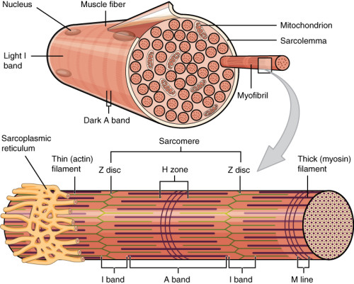
Preamble: The striated muscle cell (rhabdomyocytes) is multinucleated and rich in mitochondria. The myosin and actin proteins are responsible for contraction, under the dependence of an inflow of intracellular calcium.
A team from the University of Duke (Truskey George A) recreates a muscle unit by culturing human muscle cells taken from volunteers (1). By application of an electrical stimulus, they showed that this muscle on a chip contracts in proportion to the intensity of the current.
Moreover, they showed that the contraction passes through the release of calcium, exactly as in physiological conditions.
This short video about organs on a chip shows the spontaneous and induced rhythmic contractions of two muscles on a chip models.
A team from MIT created a neuro-muscular unit-on-a-chip (2). The neurons have been genetically engineered to respond to luminous stimuli, which once stimulated will excite the muscle cells that will contract. The rhabdomyocytes and neurons originate from mouse embryonic cells, which have been differentiated in a 3D hydrogel.
A group from the Brown Medical School Hospital (Vandenburgh H) studied Duchenne disease by reconstructing a muscle-on-a-chip from isolated myoblasts of MDX mice (genetically modified to have Duchenne myodystrophy) (3). The bio-artificial muscles are grown in 3D in a gel reproducing the extracellular matrix in a PDMS chip. This system is semi-automated, and therefore potentially compatible with high-throughput screening techniques.
Researchers from the Wyss Institute (Parker KK) developed muscle-on-a-chip applied to the tongue (4). This original approach makes possible the investigation of the muscular mechanisms involved in Duchesne’s disease. The authors showed that the muscle fibers of patients with Duchenne disease were smaller, and had a different polarization than healthy rhabdomyocytes. This results in fewer contraction capabilities of the muscles.
What's next in the muscle on a chip field?
The future in organs on a chip dedicated to muscle mainly concerns connection to other organs via the development of vessels with a contraction capacity to have the most complete vision possible of the distribution of a molecule in the human body.
References
1: Elife. 2015 Jan 9;4:e04885. doi: 10.7554/eLife.04885. Bioengineered human myobundles mimic clinical responses of skeletal muscle to drugs. Madden L, Juhas M, Kraus WE, Truskey GA, Bursac N.
2: Sci Adv. 2016 Aug 3;2(8):e1501429. doi: 10.1126/sciadv.1501429. eCollection 2016 Aug. Microfluidic device for the formation of optically excitable, three-dimensional, compartmentalized motor units. Uzel SG, Platt RJ, Subramanian V, Pearl TM, Rowlands CJ, Chan V, Boyer LA, So PT, Kamm RD
3: FASEB J. 2009 Oct; 23(10): 3325–3334. Automated drug screening with contractile muscle tissue engineered from dystrophic myoblasts. Herman Vandenburgh, Janet Shansky, Frank Benesch-Lee, Kirsten Skelly, Janelle M. Spinazzola, Yero Saponjian, and Brian S. Tseng
4: J Cell Biol. 2016 Oct 10;215(1):47-56. Epub 2016 Oct 3. A human in vitro model of Duchenne muscular dystrophy muscle formation and contractility. Nesmith AP, Wagner MA, Pasqualini FS, O’Connor BB, Pincus MJ, August PR, Parker KK.
FAQ
Skeletal muscles are a large part of human body weight, accounting for 40%, and they use about 30% of energy intake. A large loss of muscle mass often accompanies chronic pathologies, which can worsen a patient’s general condition. Curative treatments are not yet available for most musculoskeletal disorders. For example, diseases such as Duchenne’s disease and amyotrophic lateral sclerosis still lack effective treatment. Also, some treatments for chronic conditions like diabetes have undesired effects on the striated muscles. Access to a "muscle on a chip" would permit study into these adverse effects. It would also allow for a better prediction of a drug candidate’s effects during the clinical phase.
The striated muscle cell is also known as a rhabdomyocyte. This type of cell is multinucleated. It is also described as being rich in mitochondria. The proteins responsible for contraction are myosin and actin. This contraction process is dependent on an inflow of intracellular calcium. These are the basic cellular properties. Understanding them is necessary for building functional muscle models. These models are used in research. For instance, recreating the calcium-dependent contraction is a central validation step for a "muscle on a chip".
A muscle unit was recreated by one team from Duke University. This was done by culturing human muscle cells obtained from volunteers. An electrical stimulus was applied to this "muscle on a chip". It was shown that the muscle contracts in proportion to the intensity of the current that was applied. This demonstrates a controlled response. It was also found that the contraction passes through the release of calcium. This mechanism is exactly the same as what is observed in physiological conditions. Another team from MIT created a neuro-muscular unit on a chip. In this model, engineered neurons respond to light stimuli. The stimulated neurons then excite the muscle cells, causing them to contract. This shows a functional connection between nerve and muscle cells in the chip model.
Models of "muscle on a chip" have been applied to study Duchenne disease. One group from Brown Medical School Hospital reconstructed a model using myoblasts from MDX mice. These mice are genetically modified to have Duchenne myodystrophy. The bio-artificial muscles were grown in 3D within a gel that reproduced the extracellular matrix. This system was developed in a PDMS chip. Another original approach was developed by researchers from the Wyss Institute. They applied a "muscle on a chip" to the tongue. This model was also used to study the mechanisms of Duchenne’s disease. It was shown by the authors that muscle fibres from patients with this disease were smaller. They also had a different polarization compared to healthy rhabdomyocytes, which results in fewer contraction capabilities.
The future for "organs on a chip" that are dedicated to muscle is described as concerning the connection to other organs. This would be accomplished by the creation of vessels that have a contraction capacity. The objective of this work is to have the most complete vision possible of how a molecule is distributed in the human body. This integration would allow for more complex studies. Such studies would better replicate the systemic interactions that occur within a living organism. This would represent a move from isolated muscle models to interconnected organ systems.