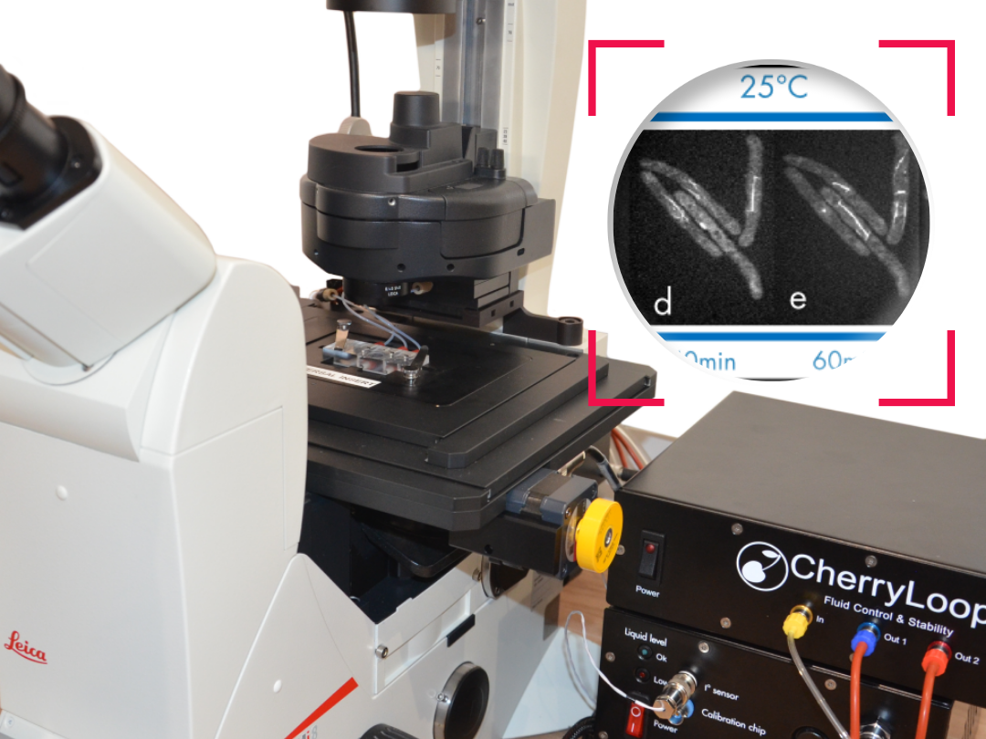
Our temperature controller is the most precise, stable and homogeneous device to make sure the targeted temperature input is the actual temperature at your sample location.
In 2014, R. Cole reviewed live-cell imaging and pointed out some key aspects such as temperature control.
Below is a snippet of his paper:
Temperature-happy and homeostatic cells
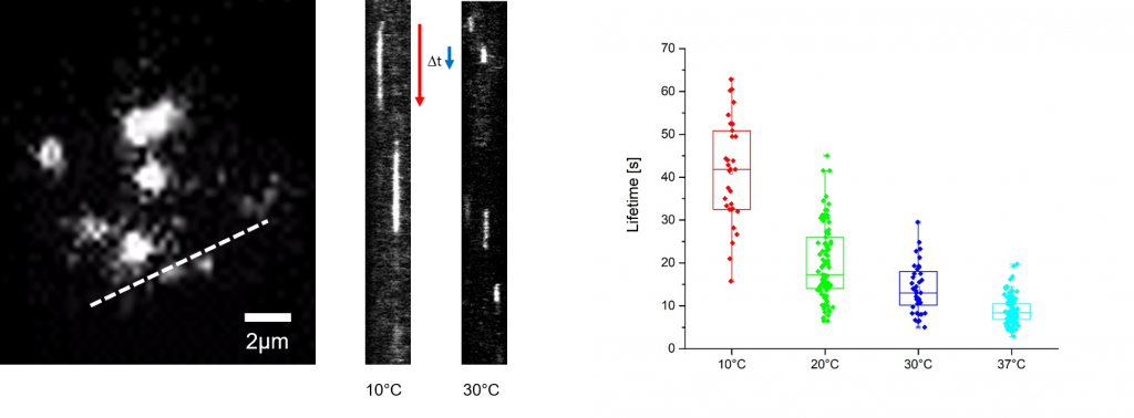
While maintaining homeostatic temperatures during imaging may seem like a no-brainer, the actual temperature of the cells being imaged is often lower than anticipated. So does a couple of degrees really matter? Yes, many cells types will stall and even reverse the cell cycle at 20–22°C,44 see Figure 4. At temperatures between 23 and 35, the timing of mitosis is dilated indicating a substantial stress on the cells. One of the main reasons for lower temperatures than expected is the use of immersion objective lenses without the use of an objective lens heater. The microscope itself acts as a massive heat sink, causing as much as a 10°C temperature differential. Even with dry objectives there can be a significant temperature differential between the edges of the specimen holder and where the cells are being imaged. It is therefore imperative that the temperature where the cells are growing be measured not just the chamber holding them. This can be accomplished with an inexpensive non-contact thermometer equipped w/laser sighting, such as OS-PAL (∼$55.00 from OMEGA Engineering, INC.). When using immersion objectives or where there is not a line of sight to the imaging area (e.g., inverted microscopes), a small thermocouple probe can be directly inserted at the focal point of the microscope. This can be safely done by attaching a small probe to the coverslip (where the cells would normally be attached) and assembling the chamber normally, including filling with media or water. Depending on the chamber type, a small hole may need to be drilled to feed the probe wire through.
One side-effect of heating above ambient temperature is stage drift. Microscopes are constructed mostly of metal with little or no engineered thermal breaks. This is especially problematic with stage type heaters but can also affect other types. To minimize the effect of temperature, allow the entire system to equilibrate (∼45–60 min), including a mock specimen chamber and focused objective lens (if immersion). While this will help, it will not be enough to maintain the same focal plane for long-term filming. The good news is that all the microscope manufacturers offer some sort of focal correction that will maintain the same focal plane indefinitely.
References
Cole R., Live-Cell Imaging, Cell Adh. Migr., 2014
How to culture vascularized & immunocompetent 3D models in a standard Multiwell
FAQ
Maintaining homeostatic temperatures is a necessary consideration for live imaging. The actual temperature of the cells being imaged is often lower than what is anticipated. It is important to correct this because even a difference of a couple of degrees matters. For example, many cell types will stall or even reverse the cell cycle when their temperature is 20–22°C. Furthermore, at temperatures between 23°C and 35°C, the timing of mitosis is dilated. This dilation indicates that a substantial stress is being placed on the cells, altering their normal function.
A main reason for lower-than-expected temperatures is the use of immersion objective lenses, particularly when they are used without an objective lens heater. The microscope itself acts as a large heat sink. This effect can cause a temperature differential of as much as 10°C between the chamber and the cells. Even when using dry objectives, a notable temperature differential can still exist. This difference in temperature is often observed between the edges of the specimen holder and the specific central area where the cells are actually being imaged.
It is necessary that the temperature is measured directly where the cells are growing, not just in the chamber holding them. This measurement can be accomplished with an inexpensive non-contact thermometer that is equipped with laser sighting, such as an OS-PAL. When using immersion objectives or on inverted microscopes where there is no line of sight, a different method is needed. A small thermocouple probe can be directly inserted at the focal point of the microscope. This can be safely done by attaching the small probe to the coverslip, where cells would normally be, and assembling the chamber as usual, including filling it with media or water.
Stage drift is a side-effect that can occur as a result of heating the microscope system above the ambient temperature. Microscopes are constructed mostly of metal components. These metal structures typically have little or no engineered thermal breaks. Because of this construction, the microscope acts as a large heat sink. This becomes especially problematic with stage-type heaters, although it can also affect other heating types. The change in temperature causes the metal to expand or contract, which results in a slow “drift” of the stage and the sample, moving it out of focus.
To minimise the effect of temperature-related drift, the entire system should be allowed to equilibrate. This process should take approximately 45 to 60 minutes. The equilibration setup should include a mock specimen chamber and have the objective lens (if using immersion) in place and focused. While this procedure will help, it will not be sufficient to maintain the same focal plane for long-term filming. To solve this, all microscope manufacturers offer some type of focal correction. These systems are able to maintain the same focal plane for extended periods.