Introduction
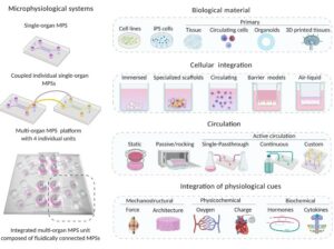
The Organ-on-a-Chip (OOAC) is a leading organ biomimetic (POB) device based on a microfluidic chip in the list of the top ten innovative technologies. The microenvironment of the chip simulates the organ in terms of tissue interfaces and mechanical stimulation by combining cell biology, engineering, and biomaterial science.
The structural and functional features of human tissue are reflected and a series of factors including drug reactions and environmental effects can be predicted in response. It is expected that with the rising of a new complex in vitro model (CIVM) and micro physiological system organ-on-chip (liver model), and particularly liver on-chip will become prominent during the drug development phases, fundamental research as well as personalized medicine.
This paper from Polidoro et al. recapitulates all the work done until now, and future perspectives.
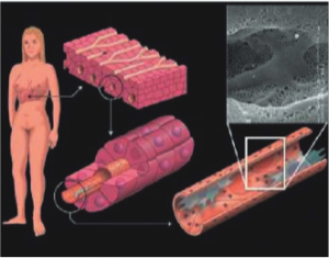
How to culture vascularized & immunocompetent 3D models in a standard Multiwell
Abstract
The authors state “The liver is one of the most studied organs of the human body owing to its central role in xenobiotic and drug metabolism. In recent decades, extensive research has aimed at developing in vitro liver models able to mimic liver functions to study pathophysiological clues in high-throughput and reproducible environments. Two-dimensional (2D) models have been widely used in screening potential toxic compounds but have failed to accurately reproduce the three-dimensionality (3D) of the liver milieu.
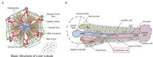
To overcome these limitations, improved 3D culture techniques have been developed to recapitulate the hepatic native microenvironment. These models focus on reproducing the liver architecture, representing both parenchymal and non-parenchymal cells, as well as cell interactions.
More recently, Liver-on-Chip (LoC) models have been developed with the aim of providing physiological fluid flow and thus achieving essential hepatic functions. Given their unprecedented ability to recapitulate critical features of the liver cellular environments, LoC has been extensively adopted in pathophysiological modeling and currently represents a promising tool for tissue engineering and drug screening applications.
In this review, we discuss the evolution of experimental liver models, from the ancient 2D hepatocyte models, widely used for liver toxicity screening, to 3D and LoC culture strategies adopted for mirroring a more physiological microenvironment for the study of liver diseases.”
References
Anna Polidoro M, Ferrari E, Marzorati S, Lleo A, Raponi M. Experimental liver models: from cell culture techniques to microfluidic organs-on-chip. Liver Int. 2021 May 9. doi: 10.1111/liv.14942. Epub ahead of print. PMID: 33966344.
FAQ
The liver is a frequently studied organ. Extensive research has sought to create laboratory models of the liver. For a long time, flat, two-dimensional models were widely used. These models were applied to screening compounds for toxicity. A failure of these flat models was identified. They were unable to accurately show the three-dimensional nature of the liver’s surroundings. Because of this limitation, improved culture techniques were needed. These new methods were developed to better represent the native hepatic environment. The focus of these newer models is on the liver’s architecture and cellular interactions.
Certain new technologies are being developed to copy organ functions. These devices are based on chips that use small fluid channels. The environment of the organ is recreated on the chip. This includes representing tissue interfaces and mechanical forces. Several fields are combined to make these devices, including cell biology, engineering, and biomaterial science. The structural and functional characteristics of human tissue are shown in the model. A series of factors, such as reactions to compounds and environmental effects, can be forecasted. These systems are expected to become prominent in research and medicine.
Improved three-dimensional culture techniques were developed to show the hepatic environment. The focus of these models is placed on representing the liver’s architecture. This includes both parenchymal and non-parenchymal cells, as well as their interactions. More recently, specific liver models on chips have been created. These systems were designed with the aim of providing fluid flow as it occurs in the body. Through this fluid motion, important hepatic functions are achieved. These chip-based liver models are now widely applied for modelling disease processes. They are also considered a useful tool for tissue engineering and compound-testing applications.
The liver is one of the most studied organs in the body. This is because of its main function in the metabolism of foreign substances and medications. For a long time, extensive research has been aimed at developing laboratory liver models. These models are desired to be able to copy liver functions. They are used to study clues related to disease processes. An environment that allows for repeatable experiments is also a goal. While flat models were widely used for screening liver toxicity, newer strategies are now adopted. These newer three-dimensional and chip-based cultures are used to provide a more representative setting for the study of liver diseases.

