Temperature control of microtubules assembly in mammalian cells
We developed a temperature control heater/cooler device that answers microscope imaging needs for any type of cells. Our system features an ultra fast temperature shift: 5 to 45C in seconds to circumvent thermal gradient-generated artifacts, and microfluidic technology to prevent fluid flow shear stress on the sample. We specifically designed it to reach below ambient temperature to allow cytoskeleton, endocytosis and high-resolution imaging studies. We made sure that the system is plug and play and fits on any microscope setting.
Mammalian cells microtubules disassembly
At the cellular level, many processes are temperature regulated. For instance, incubating cells on ice for 30 min is sufficient to induce microtubules depolymerization. At 19C, endocytosis is arrested and at lower temperature vesicular transport is blocked. Protein aggregation can also be induced with low or high temperatures. Moreover temperature-sensitive spontaneous mutations or engineered mutations have allowed to specifically control any genes of interest. All of these processes are very fast acting, and combining them with live cell imaging requires a device that can change temperature very quickly, that is robust, and respect cell integrity by preventing shear stress. Here, we present the development and validation of CherryTemp, a temperature controller for cell biology microscope imaging.
Microtubule cold-induced experiments
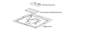
1. Temperature controller device. Our device is using microfluidic technology with a PDMS chip composed of microchannels allowing the circulation of thermalized fluid. The chip is crosslinked to a coverslip and is connected to Peltier devices set at two different hot/cold temperatures making possible to quickly change from low to high temperatures. Our Cherrybox electronically controls the Peltier element. In our system, the fluid is never in contact with the sample, preventing any shear stress.
2. Cell culture. GFP-Atb2 (tubulin) expressing HeLa cells were plated on glass coverslides onto which we added biocompatible tape to delimit the cultured area. Cells were cultured with DMEM 10%FBS, at 37°C, with 5% CO2. The day of the experiment, coverslides were harvested, and one drop of medium was added onto the cells, we then simply place the Cherrytemp chip/coverslip onto them and started temperature shift experiment.
3. Microscope imaging. Temperature shift experiments were done using an inverted Nikon TE2000e microscope and a 100/1.45 oil immersion objective. Images were acquired using Metamorph 7.5 software for microscope and Hamamatsu CCD camera control.
Effective and reversible microtubules depolymerization upon cold-induction
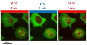
We used GFP-Atb2 expressing HeLa cells to track microtubules depolymerization. The field of observation was chosen to have a representative aspect of microtubule network in interphase and in mitosis. At 37C, we observed spider-like microtubules network, which elongates from the MTOC. We also observed a mitotic cell with typical metaphasic spindles. Upon shifting temperature at 5C for 6 min, we induced complete microtubules depolymerization. This was characterized by a drop in microtubules-associated fluorescence and an increase in cytoplasmic fluorescence. Indicating that tubulin dimers depolymerized and filled the pool of free cytoplasmic tubulin. When we shifted the temperature back to 37C, microtubules network re-polymerized demonstrating the reversibility of this process.

Ultra fast temperature shift device for in vitro experiments under microscopy
Temperature regulation of mammalian cells microtubules stability
Fast, reversible and below ambient
CherryTemp enables scientists to shift temperature from 5 to 45C in 10 seconds. Existing temperature controller can take minutes (incubation box, microscope stages) to change temperature generating thermal gradient on the sample (>3C). The quick action of our temperature controller allows scientists to induce and revert cellular phenotypes. We showed that we could fully depolymerize and repolymerize microtubules network within minutes. With our system, cell biologists can also slowly increment temperature and generate hypomorphic-phenotypes. Developing a system that could reach below ambient temperature was an important requirement to enable to study vesicular transport, cytoskeleton or to perform high-resolution imaging.
Temperature-control for mammalian cells microscopy
Temperature control intervenes in many cellular processes. With CherryTemp one can cold-induced endocytosis arrest, stop all vesicular transport and trafficking, induce protein recombination and track it with live-cell imaging, record the first steps of stem cells differentiation, induce and revert protein aggregation, induce heat-gated ion channel, track virus entry and trafficking inside the cells with microscope imaging.
References
Jeremy Cramer, study and characterization of a heater/cooler device: temperature control for cell biology, Master thesis, 2012.
S. Ben-Aroya, X. Pan, JD. Boeke, and P. Hieter, Making temperature-sensitive mutants, Methods Enzymol. 2010
FAQ
Microtubules in mammalian cells are known to be regulated by temperature. This process has been shown to be reversible. For example, in experiments using HeLa cells that express GFP-Atb2 (tubulin), a spider-like network of microtubules is observed at 37°C. This network elongates from the microtubule organising centre. When these cells are subjected to a temperature of 5°C for six minutes, complete depolymerization of the microtubules is induced. This change is seen as a decrease in fluorescence associated with the microtubules. An increase in fluorescence in the cytoplasm is also seen, which indicates the tubulin dimers have depolymerized into the free cytoplasmic pool. When the temperature is returned to 37°C, the microtubule network re-polymerizes.
Besides microtubule assembly, many processes at the cellular level are regulated by temperature. It has been noted that endocytosis is arrested at 19°C. At temperatures lower than this, vesicular transport is blocked. The aggregation of proteins can also be induced by exposure to either low or high temperatures. This effect can also be reversed. Other areas of study that involve temperature control include endocytosis arrest and the complete stop of vesicular transport and trafficking. Temperature-gated ion channels can be induced. Protein recombination can be started and then tracked. The first steps of stem cell differentiation can also be recorded. Additionally, virus entry and its subsequent trafficking inside cells can be followed with microscope imaging.
An experiment was conducted using HeLa cells that express GFP-Atb2, which is a tubulin marker. These cells were plated on glass coverslides. Biocompatible tape was added to the coverslides to delimit the cultured area. The cells were cultured in DMEM with 10% FBS at 37°C and 5% CO2. On the day of the experiment, the coverslides were harvested. A drop of medium was added to the cells. A specific temperature control chip and coverslip system was then placed onto the cells. The experiment was performed using an inverted Nikon TE2000e microscope. A 100/1.45 oil immersion objective was used for observation. Images were acquired using Metamorph 7.5 software to control the microscope and a Hamamatsu CCD camera.
The described temperature controller device uses microfluidic technology. It is designed to prevent fluid flow shear stress from affecting the sample. The system is based on a PDMS chip. This chip contains microchannels, which permit the circulation of a thermalized fluid. The chip is crosslinked to a coverslip. It is also connected to Peltier devices that are set at two different hot and cold temperatures. This setup makes it possible to quickly change from low to high temperatures. An electronic controller manages the Peltier elements. In this specific system, the thermalized fluid circulates within the chip’s microchannels. The fluid is never in direct contact with the biological sample. This separation is what prevents any shear stress from being applied to the cells during the temperature change.