Brain: No-human in-vivo model
The human brain is one of the most convoluted organs. Contemporaneously, in-vivo and in-vitro (organotypic brain cultures for example) non-human models are unraveling the mechanism of the complex brain signaling and additionally are being used to identify the therapeutic agents.
Several vertebrates including zebrafish, mice, and rat are applied to prototype human neurodegeneration. Neuro-disorders are either replicated chemically or genetically imitated through mutation in selected no-human models. However, many drug contenders failed to meet the translation from no-human to human model after the effective screening in the animal. Often, no-human models partially exhibit the etiology and symptomatic traits of stimulated or mutated diseases. As the anatomy, physiology, and biochemistry of the human brain are substantially different from the modeled vertebrates, henceforth the comparison concerning no-human and human model is impractical.
How to culture vascularized & immunocompetent 3D models in a standard Multiwell
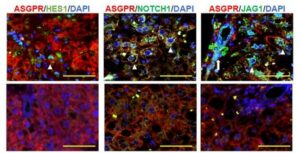
In-vitro analysis of human brain cells: clinical applications
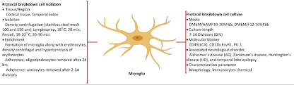
Generally, in-vitro humanmodels encompass the individual or mixed culture of primary brain cells separated from different age human or preserved cell lines. Human brain cell culturing involves rigorous and laborious biological protocols. The protocols have been devised in two phases: 1) protocol cell isolation includes the cell dissociation measures from sliced tissue, 2) protocol cell culture relates to the developing etiquettes for the considered neurological disorder. Referenced studies correlated with brain cell culture are being utilized to unveil the processes including inflammation, neurotoxicity, and neuroprotection, additionally, to screen for pre-clinical drugs to treat neurodegenerative disorders.
Limitations of dissociate human culture compensated with slice culture
The chief disadvantage of the dissociate culture is associated with brain anatomical and cellular interactions, which cannot be replicated easily. Moreover, the number of autopsies is declined day by day; hence, the availability of the brain tissue is limited. In this scenario, more often, researchers find that the yield of brain cells is relatively low after tissue dissociation.
In contrast, the advancement in slice culture method enables limited anatomical and cellular interactions and offers a model nearer to the in vivo situation. Additionally, Verwer 2002 claims that brain tissue obtained from an autopsy can be retained for a more extended period (up to 78 days), and further can be manipulated.
Organotypic brain cultures: Apprehension from upcoming technologies
Further modification in slice culture method will deliver a robust system for analyzing the fundamental biology of human brain cells and their wirings. The system will enable the understanding of basic procedures of brain cell death, cell repairing, and testing novel protocols for brain disorders. Slice culture can be utilized to explore electrical properties and synaptic functioning of the human brain via applying electrophysiological approaches. These cultures could be established as standard studies to unfold mechanisms of neuroinflammation, the death of human neurons, and glial scar formation.
Discover our brain project !
References
- Aderem A, and Underhill DM. Mechanisms of phagocytosis in macrophages. Annual Review of Immunology1999;17:593–623.
- Brewer GJ, et al. Culture and regeneration of human neurons after brain surgery. Journal of Neuroscience Methods 2001;107:15–23.
- Gonzalez-Martinez JA, et al. Neurogenesis in the postnatal human epileptic brain. Journal of Neurosurgery2007;107:628–35.
- Konishi Y, et al . Isolation of living neurons from human elderly brains using the immunomagnetic sorting DNA-linker system. American Journal of Pathology2002;161:1567–76.
- McKhann GM,et al. The isolation of neurons from normal and abnormal human cerebral cortex. Archives of Neurology1969;20:542–7.
- Ridet JL, et al. Transplantation of human adult astrocytes: efficiency and safety requirements for an autologous gene therapy. Journal of Neuroscience Research2003;72:704–8.
- Verwer RW, et al. Cells in human postmortem brain tissue slices remain alive for several weeks in culture. FASEB Journal2002;16:54–60.
- Walsh K, et al. Human central nervous system tissue culture: a historical review and examination of recent advances. Neurobiology of Disease2005;18:2–18.
- Wroblewska Z, et al. Human brain in tissue culture. II. Studies of long-term cultures. Journal of Comparative Neurology 1975;161:307–16
FAQ
The human brain is a very involved organ. Currently, non-human in-vivo and in-vitro models are used to understand its elaborate signalling and to identify therapeutic agents. Vertebrates like zebrafish, mice, and rats are applied to prototype human neurodegeneration. Neuro-disorders are either replicated chemically or genetically in these models. A problem exists, as many potential drugs fail when translated from the non-human to the human model. This is often because the non-human models only partially show the symptomatic traits of the diseases. The anatomy, physiology, and biochemistry of the human brain are substantially different from these vertebrates. This makes a direct comparison between non-human and human models impractical.
Generally, human in-vitro models are composed of individual or mixed cultures of brain cells. These cells can be primary cells separated from human tissue or preserved cell lines. The process for culturing human brain cells involves demanding and laborious biological protocols. These protocols are devised in two main phases. The first phase is cell isolation, which includes measures for cell dissociation from sliced tissue. The second phase, protocol cell culture, relates to the developing etiquettes for the specific neurological disorder being considered. Studies using brain cell culture are applied to unveil processes such as inflammation, neurotoxicity, and neuroprotection. They are also used to screen pre-clinical drugs for neurodegenerative disorders.
The main disadvantage of the dissociate culture method is associated with brain anatomical and cellular interactions. These features cannot be replicated easily in this type of culture. There are also practical challenges. The number of autopsies is declining, which limits the availability of brain tissue. In this situation, researchers often find that the yield of brain cells after tissue dissociation is relatively low. In contrast, the advancement in the slice culture method provides a model that is nearer to the in vivo situation. This is because it enables limited anatomical and cellular interactions to be maintained. Additionally, brain tissue from an autopsy can be retained for a more extended period, up to 78 days, and can be further manipulated.
Further modification of the slice culture method is expected to deliver a dependable system. This system will be for analyzing the fundamental biology of human brain cells and their wirings. It will enable a better understanding of the basic procedures of brain cell death and cell repairing. The testing of new protocols for brain disorders will also be possible. Slice culture can be utilized to explore the electrical properties and synaptic functioning of the human brain. This is done by applying electrophysiological approaches. It is thought these cultures could be established as standard studies. They would be used to unfold the mechanisms of neuroinflammation, the death of human neurons, and the formation of glial scars.