Abstract of mmb1p Binds Mitochondria to Dynamic Microtubules
Mitochondria form a dynamic tubular network within the cell. Proper mitochondria movement and distribution are critical for their localized function in cell metabolism, growth, and survival. In mammalian cells, mechanisms of mitochondria positioning appear dependent on the microtubule cytoskeleton, with kinesin or dynein motors carrying mitochondria as cargos and distributing them throughout the microtubule network. Interestingly, the timescale of microtubule dynamics occurs in seconds, and the timescale of mitochondria distribution occurs in minutes. How does the cell couple these two time constants?
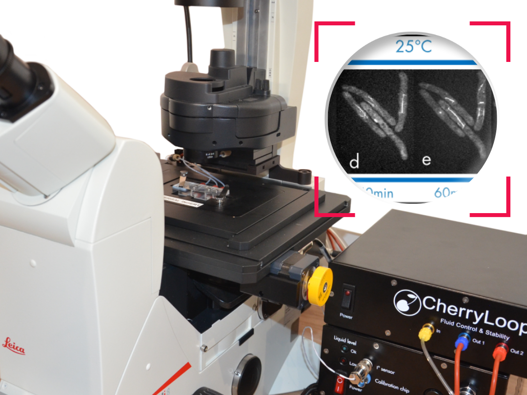
Video extracted from the publication.
How to culture vascularized & immunocompetent 3D models in a standard Multiwell
References
https://www.sciencedirect.com/science/article/pii/S0960982211007822Chuanhai Fu, Deeptee Jain, Judite Costa, Guilhem Velve-Casquillas, Phong T. Tran, mmb1p Binds Mitochondria to Dynamic Microtubules, Current Biology, Volume 21, Issue 17, 2011, Pages 1431-1439,
FAQ
The proper movement and distribution of mitochondria are considered critical. This importance is due to their localized function within the cell. These functions are necessary for cell metabolism, growth, and survival. In mammalian cells, the mechanisms for positioning mitochondria appear to be dependent on the microtubule cytoskeleton. This network is used to distribute the mitochondria. The mitochondria are carried as cargos by specific motors. These motors are identified as kinesin or dynein. They transport the mitochondria along the microtubule network, ensuring they are distributed correctly to support their localized functions.
An interesting point is raised regarding the timescales of the components involved. The dynamics of the microtubules themselves occur very quickly. This timescale is reported to be in seconds. In contrast, the distribution of the mitochondria, which uses the microtubules, happens much more slowly. The timescale for mitochondria distribution is reported to be in minutes. This difference presents a challenge. A question is posed about how the cell manages to couple these two different time constants. How the rapid, seconds-long dynamics of the microtubules are coupled to the slower, minutes-long process of distributing mitochondria is a key question.